

Theoretical Analysis and Expression Profiling of 17β-Hydroxysteroid Dehydrogenase Genes in Gonadal Development and Steroidogenesis of Leopard Coral Grouper (Plectropomus leopardus)
- PMID: 38396857
- PMCID: PMC10889806
- DOI: 10.3390/ijms25042180
Theoretical Analysis and Expression Profiling of 17β-Hydroxysteroid Dehydrogenase Genes in Gonadal Development and Steroidogenesis of Leopard Coral Grouper (Plectropomus leopardus)
Abstract
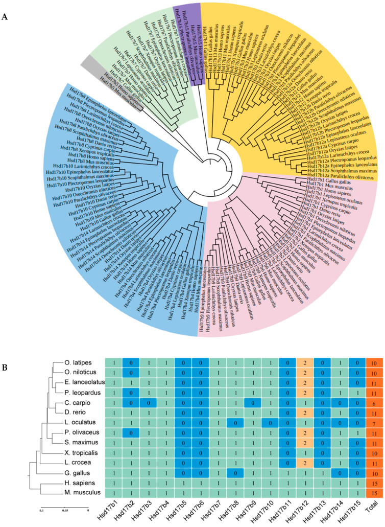
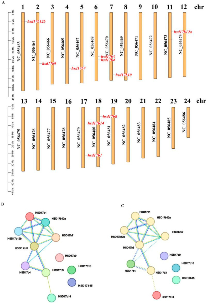
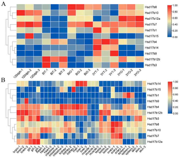
The differentiation and developmental trajectory of fish gonads, significantly important for fish breeding, culture, and production, has long been a focal point in the fields of fish genetics and developmental biology. However, the mechanism of gonadal differentiation in leopard coral grouper (Plectropomus leopardus) remains unclear. This study investigates the 17β-Hydroxysteroid Dehydrogenase (Hsd17b) gene family in P. leopardus, with a focus on gene characterization, expression profiling, and functional analysis. The results reveal that the P. leopardus's Hsd17b gene family comprises 11 members, all belonging to the SDR superfamily. The amino acid similarity is only 12.96%, but conserved motifs, such as TGxxxGxG and S-Y-K, are present in these genes. Hsd17b12a and Hsd17b12b are unique homologs in fish, and chromosomal localization has confirmed that they are not derived from different transcripts of the same gene, but rather are two independent genes. The Hsd17b family genes, predominantly expressed in the liver, heart, gills, kidneys, and gonads, are involved in synthesizing or metabolizing sex steroid hormones and neurotransmitters, with their expression patterns during gonadal development categorized into three distinct categories. Notably, Hsd17b4 and Hsd17b12a were highly expressed in the testis and ovary, respectively, suggesting their involvement in the development of reproductive cells in these organs. Fluorescence in situ hybridization (FISH) further indicated specific expression sites for these genes, with Hsd17b4 primarily expressed in germ stem cells and Hsd17b12a in oocytes. This comprehensive study provides foundational insights into the role of the Hsd17b gene family in gonadal development and steroidogenesis in P. leopardus, contributing to the broader understanding of fish reproductive biology and aquaculture breeding.
Keywords: 17β-Hydroxysteroid Dehydrogenase; Plectropomus leopardus; gonadal development; sex differentiation; steroidogenesis.
Conflict of interest statement
The authors declare no conflicts of interest.
Figures


Similar articles
-
Characteristics and sex dimorphism of 17β-hydroxysteroid dehydrogenase family genes in the olive flounder Paralichthys olivaceus.J Steroid Biochem Mol Biol. 2020 May;199:105597. doi: 10.1016/j.jsbmb.2020.105597. Epub 2020 Jan 17. J Steroid Biochem Mol Biol. 2020. PMID: 31958634
-
Genome wide analysis of the sox32 gene in germline maintenance and differentiation in leopard coral grouper (Plectropomus leopardus).Comp Biochem Physiol Part D Genomics Proteomics. 2025 Jun;54:101402. doi: 10.1016/j.cbd.2024.101402. Epub 2024 Dec 28. Comp Biochem Physiol Part D Genomics Proteomics. 2025. PMID: 39742679
-
Characterization of eight types of 17β-hydroxysteroid dehydrogenases from the Japanese sardine Sardinops melanostictus: The probable role of type 12a in ovarian estradiol synthesis.Gen Comp Endocrinol. 2024 Feb 1;347:114423. doi: 10.1016/j.ygcen.2023.114423. Epub 2023 Dec 10. Gen Comp Endocrinol. 2024. PMID: 38086427
-
Synchronously sexual maturity in hermaphrodite fish as revealed by transcriptome analysis in Plectropomus leopardus.Gene. 2024 Apr 5;901:148166. doi: 10.1016/j.gene.2024.148166. Epub 2024 Jan 17. Gene. 2024. PMID: 38242379
-
A working hypothesis for the regulation of steroidogenesis and germ cell development in the gonads by glucocorticoids and 11 beta-hydroxysteroid dehydrogenase (11 beta HSD).Mol Cell Endocrinol. 1994 Apr;100(1-2):55-63. doi: 10.1016/0303-7207(94)90279-8. Mol Cell Endocrinol. 1994. PMID: 8056159 Review.
Cited by
-
Genomic Signatures of Domestication in European Seabass (Dicentrarchus labrax L.) Reveal a Potential Role for Epigenetic Regulation in Adaptation to Captivity.Ecol Evol. 2024 Dec 3;14(12):e70512. doi: 10.1002/ece3.70512. eCollection 2024 Dec. Ecol Evol. 2024. PMID: 39629177 Free PMC article.
References
-
- Butka E.G., Freedberg S. Population structure leads to male-biased population sex ratios under environmental sex determination, Evolution. Int. J. Org. Evol. 2019;73:99–110. - PubMed
-
- Devlin R.H., Nagahama Y. Sex determination and sex differentiation in fish: An overview of genetic, physiological, and environmental influences. Aquaculture. 2002;208:191–364. doi: 10.1016/S0044-8486(02)00057-1. - DOI
-
- Nakamoto M., Shibata Y., Ohno K., Usami T., Kamei Y., Taniguchi Y., Todo T., Sakamoto T., Young G., Swanson P., et al. Ovarian aromatase loss-of-function mutant medaka undergo ovary degeneration and partial female-to-male sex reversal after puberty. Mol. Cell. Endocrinol. 2018;460:104–122. doi: 10.1016/j.mce.2017.07.013. - DOI - PubMed
MeSH terms
Substances
Grants and funding
- ZDYF2023XDNY182/the Key Research and Development Project of Hainan Province
- 2022ZLGX01/the Key Research and Development Project of Shandong Province
- No. 32202927/the National Natural Science Foundation of China
- HSPHDSRF-2023-02-005/the PhD Scientific Research and Innovation Foundation of Sanya Yazhou Bay Science and Technology City
LinkOut - more resources
Full Text Sources
Miscellaneous
