. 2024 Feb 12;25(4):2210.
doi: 10.3390/ijms25042210.

Evaluating the Effects of Kidney Preservation at 10 °C with Hemopure and Sodium Thiosulfate in a Rat Model of Syngeneic Orthotopic Kidney Transplantation

Affiliations

Evaluating the Effects of Kidney Preservation at 10 °C with Hemopure and Sodium Thiosulfate in a Rat Model of Syngeneic Orthotopic Kidney Transplantation

Maria Abou Taka et al. Int J Mol Sci. .

Abstract

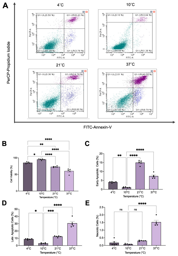
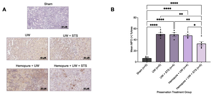
Kidney transplantation is preferred for end-stage renal disease. The current gold standard for kidney preservation is static cold storage (SCS) at 4 °C. However, SCS contributes to renal graft damage through ischemia-reperfusion injury (IRI). We previously reported renal graft protection after SCS with a hydrogen sulfide donor, sodium thiosulfate (STS), at 4 °C. Therefore, this study aims to investigate whether SCS at 10 °C with STS and Hemopure (blood substitute), will provide similar protection. Using in vitro model of IRI, we subjected rat renal proximal tubular epithelial cells to hypoxia-reoxygenation for 24 h at 10 °C with or without STS and measured cell viability. In vivo, we preserved 36 donor kidneys of Lewis rats for 24 h in a preservation solution at 10 °C supplemented with STS, Hemopure, or both followed by transplantation. Tissue damage and recipient graft function parameters, including serum creatinine, blood urea nitrogen, urine osmolality, and glomerular filtration rate (GFR), were evaluated. STS-treated proximal tubular epithelial cells exhibited enhanced viability at 10 °C compared with untreated control cells (p < 0.05). Also, STS and Hemopure improved renal graft function compared with control grafts (p < 0.05) in the early time period after the transplant, but long-term function did not reach significance. Overall, renal graft preservation at 10 °C with STS and Hemopure supplementation has the potential to enhance graft function and reduce kidney damage, suggesting a novel approach to reducing IRI and post-transplant complications.

Keywords: Hemopure; graft and recipient survival; ischemia–reperfusion injury (IRI); kidney transplantation; sodium thiosulfate (STS); static cold storage (SCS).


Conflict of interest statement

The authors declare no conflicts of interest.

Figures

Figure 1
Figure 1
In vitro model of rat renal IRI. A summary of the in vitro rat renal IRI model used to determine the protective effects of STS at various temperatures (4 °C, 10 °C, 21 °C, and 37 °C). Figure prepared with BioRender (biorender.com). Accessed on 1 March 2023.
Figure 2
Figure 2
In vivo syngeneic model of renal IRI in a rat kidney transplantation model at 10 °C. A summary of the experimental procedure used to determine renal graft outcomes following prolonged kidney storage for 24 h at 10 °C with UW, UW with 150 µM STS, Hemopure with UW, and Hemopure with UW and 150 µM STS. Figure prepared with BioRender (biorender.com). Accessed on 1 March 2023.
Figure 3
Figure 3
The 10 °C STS treatment improved rat proximal tubule epithelial cell survival during renal IRI. (A) Representative flow cytometry images for all temperatures investigated. (B) Mean cell viability (%) (n = 5) as determined by the proportion of cells negative for FITC–Annexin-V and PerCP-PI staining. (C) Mean early apoptosis (%) (n = 5) as determined by the percentage of cells positively stained for FITC–Annexin-V and negatively stained for PerCP-PI. (D) Mean late apoptosis (%) (n = 5) as determined by the percentage of cells positively stained for FITC–Annexin-V and PerCP-PI. (E) Mean necrosis (%) (n = 5) as determined by the percentage of cells negatively stained for FITC–Annexin-V and positively stained for PerCP-PI. Lines indicate mean ± SEM. Means were analyzed using one-way ANOVA and Tukey’s post hoc test. *, p < 0.05. **, p < 0.01. ***, p < 0.001. ****, p < 0.0001. ns, not significant (p > 0.05).
Figure 4
Figure 4
The 10 °C STS treatment improved rat proximal tubule epithelial cell viability during renal IRI compared with control cells. Mean cell viability (%) for serum-treated cells (positive control; n = 5), serum-free (SF) treated cells (negative control; n = 5), and STS-treated cells (n = 5) as determined by the proportion of cells negative for FITC–Annexin-V and PerCP-PI staining. Lines indicate mean ± SEM. Means were analyzed using one-way ANOVA and Tukey’s post hoc test. *, p < 0.05. **, p < 0.01. ***, p < 0.001. ****, p < 0.0001. ns, not significant (p > 0.05).
Figure 5
Figure 5
Renal graft preservation at 10 °C modulated recipient survival following transplantation. Survival rates of kidney transplant recipients whose donor kidneys were perfused and stored for 24 h at 10 °C in UW (n = 5), UW + 150 µM STS (n = 4), Hemopure + UW (n = 4), or Hemopure + UW + 150 µM STS (n = 5) as well as those of Sham-operated rats. Survival data analyzed via Kaplan–Meier survival analysis and log-rank test. Note: one UW rat and one Hemopure + UW + STS rat survived until POD 14.
Figure 6
Figure 6
Renal graft preservation at 10 °C with Hemopure and STS improved urine osmolality. Urine osmolality levels of renal grafts preserved for 24 h at 10°C in UW (n = 5), UW + 150 µM STS (n = 4), Hemopure + UW (n = 4), or Hemopure + UW + 150 µM STS (n = 5). Osmolality levels were measured using the Model 3320 Osmometer and normalized to company-provided standards. Bars represent mean ± SEM. Values were compared using one-way ANOVA followed by Tukey’s post hoc test. *, p < 0.05. **, p < 0.01. ****, p < 0.0001.
Figure 7
Figure 7
Renal graft preservation at 10 °C with Hemopure and STS enhanced recipient kidney function following transplantation. Serum creatinine (A), blood urea nitrogen (B), and estimated glomerular filtration rate (C) of kidney transplant recipients whose donor kidneys were perfused and stored for 24 h at 10 °C in UW (n = 5), UW + 150 µM STS (n = 4), Hemopure + UW (n = 4), or Hemopure + UW + 150 µM STS (n = 5), as well as those of Sham-operated rats. Values were analyzed via one-way ANOVA and Tukey’s post hoc test. *, p < 0.05. **, p < 0.01. ***, p < 0.001. ****, p < 0.0001.
Figure 8
Figure 8
Renal graft preservation at 10 °C with Hemopure and STS reduced acute tubular necrosis after 24 h of storage. (A) Representative H&E images of formalin-fixed kidney sections on POD 3. Images were taken at 20× magnification (scale bar = 50 µM). (B) Acute tubular necrosis (ATN) scores assigned by two blinded renal pathologists (1 = <11%, 2 = 11–24%, 3 = 25–45%, 4 = 46–75%, 5 = >75%). Each individual data point indicates the score assigned to one rat kidney sample. Lines represent mean ± SEM. Values were compared using one-way ANOVA followed by Tukey’s post hoc test. *, p < 0.05. **, p < 0.01. ****, p < 0.0001.
Figure 9
Figure 9
Renal graft preservation at 10 °C with Hemopure and STS reduced apoptotic tissue injury after 24 h of storage. (A) Representative TUNEL images of formalin-fixed kidney sections on POD 3. Images were taken at 20× magnification (scale bar = 50 µM). (B) Mean %TUNEL + area determined with ImageJ v.1.53 using a ratio of TUNEL + area (brown) to total tubular area. Each individual data point indicates the mean %TUNEL + area of 10 random fields of view of one rat kidney sample. Lines represent mean ± SEM. Values were compared using one-way ANOVA followed by Tukey’s post hoc test. **, p < 0.01. ****, p < 0.0001.
Figure 10
Figure 10
Renal graft preservation at 10 °C with Hemopure and STS mitigated kidney tissue injury after 24 h of storage. (A) Representative immunohistochemical images of formalin-fixed kidney sections on POD 3 stained with Kim-1, a biomarker of renal injury. Images were taken at 20× magnification (scale bar = 50 µM). (B) Mean %KIM-1+ area determined with ImageJ using a ratio of KIM-1+ area (brown) to total tubular area. Each individual data point indicates the mean %KIM-1+ area of 10 random fields of view of one rat kidney sample. Lines represent mean ± SEM. Values were compared using one-way ANOVA followed by Tukey’s post hoc test. *, p < 0.05. **, p < 0.01. ****, p < 0.0001.
Figure 11
Figure 11
Renal graft preservation at 10 °C with Hemopure and STS reduced macrophage infiltration after 24 h of storage. (A) Representative immunohistochemical images of formalin-fixed kidney sections on POD 3 stained with CD68, a macrophage marker. Images were taken at 20× magnification (scale bar = 50 µM). (B) Mean %CD68+ area determined with ImageJ using a ratio of CD68+ area (brown) to total tubular area. Each individual data point indicates the mean %CD68+ area of 10 random fields of view of one rat kidney sample. Lines represent mean ± SEM. Values were compared using one-way ANOVA followed by Tukey’s post hoc test. *, p < 0.05. **, p < 0.01. ***, p < 0.001. ****, p < 0.0001.
Figure 12
Figure 12
Renal graft preservation at 10 °C with Hemopure and STS decreased neutrophil infiltration after 24 h of storage. (A) Representative immunohistochemical images of formalin-fixed kidney sections on POD 3 stained with MPO, which is associated with neutrophil expression. Images were taken at 20× magnification (scale bar = 50 µM). (B) Mean %MPO+ area determined with ImageJ using a ratio of MPO+ area (brown) to total tubular area. Each individual data point indicates the mean %MPO+ area of 10 random fields of view of one rat kidney sample. Lines represent mean ± SEM. Values were compared using one-way ANOVA followed by Tukey’s post hoc test. *, p < 0.05. **, p < 0.01. ****, p < 0.0001.

Similar articles

Cited by

References

    1. Thurlow J.S., Joshi M., Yan G., Norris K.C., Agodoa L.Y., Yuan C.M., Nee R. Global Epidemiology of End-Stage Kidney Disease and Disparities in Kidney Replacement Therapy. Am. J. Nephrol. 2021;52:98–107. doi: 10.1159/000514550. - DOI - PMC - PubMed
    1. Deng Y., Li N., Wu Y., Wang M., Yang S., Zheng Y., Deng X., Xiang D., Zhu Y., Xu P., et al. Global, Regional, and National Burden of Diabetes-Related Chronic Kidney Disease From 1990 to 2019. Front. Endocrinol. 2021;12:672350. doi: 10.3389/fendo.2021.672350. - DOI - PMC - PubMed
    1. Lacroix J.D., Mahoney J.E., Knoll G.A. Renal transplantation using non-heart-beating donors: A potential solution to the organ donor shortage in Canada. Can. J. Surg. 2004;47:10–14. - PMC - PubMed
    1. Himmelfarb J., Vanholder R., Mehrotra R., Tonelli M. The current and future landscape of dialysis. Nat. Rev. Nephrol. 2020;16:573–585. doi: 10.1038/s41581-020-0315-4. - DOI - PMC - PubMed
    1. Hariharan S., Israni A.K., Danovitch G. Long-Term Survival after Kidney Transplantation. N. Engl. J. Med. 2021;385:729–743. doi: 10.1056/NEJMra2014530. - DOI - PubMed