. 2024 Feb 13;25(4):2240.
doi: 10.3390/ijms25042240.

An Unusual Two-Domain Thyropin from Tick Saliva: NMR Solution Structure and Highly Selective Inhibition of Cysteine Cathepsins Modulated by Glycosaminoglycans

Affiliations

An Unusual Two-Domain Thyropin from Tick Saliva: NMR Solution Structure and Highly Selective Inhibition of Cysteine Cathepsins Modulated by Glycosaminoglycans

Zuzana Matoušková et al. Int J Mol Sci. .

Abstract

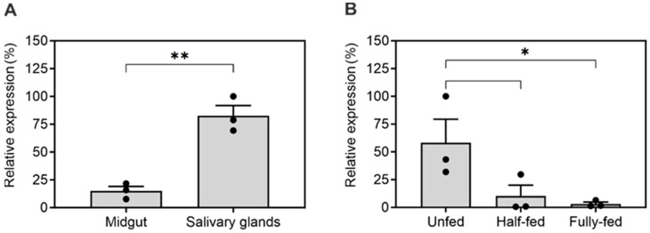
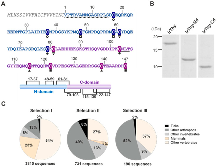
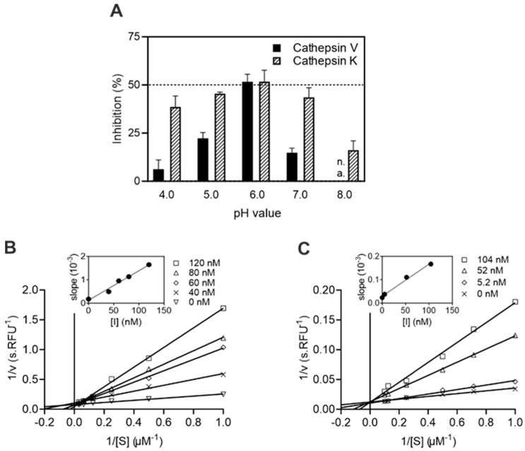
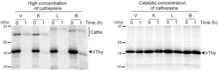
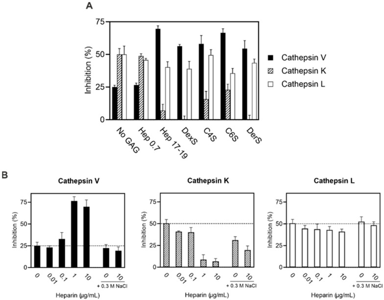
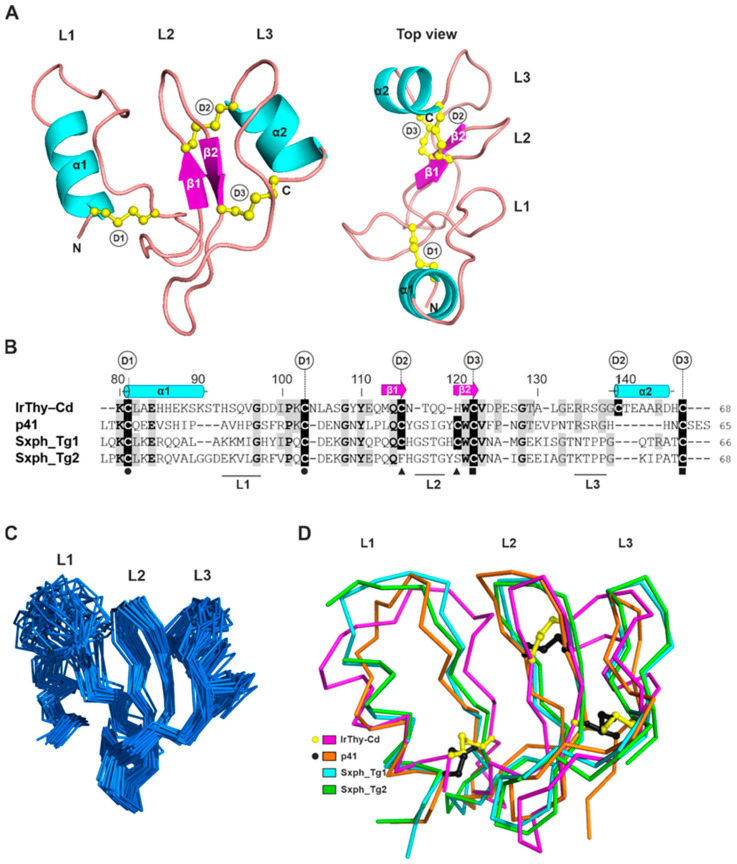
The structure and biochemical properties of protease inhibitors from the thyropin family are poorly understood in parasites and pathogens. Here, we introduce a novel family member, Ir-thyropin (IrThy), which is secreted in the saliva of Ixodes ricinus ticks, vectors of Lyme borreliosis and tick-borne encephalitis. The IrThy molecule consists of two consecutive thyroglobulin type-1 (Tg1) domains with an unusual disulfide pattern. Recombinant IrThy was found to inhibit human host-derived cathepsin proteases with a high specificity for cathepsins V, K, and L among a wide range of screened cathepsins exhibiting diverse endo- and exopeptidase activities. Both Tg1 domains displayed inhibitory activities, but with distinct specificity profiles. We determined the spatial structure of one of the Tg1 domains by solution NMR spectroscopy and described its reactive center to elucidate the unique inhibitory specificity. Furthermore, we found that the inhibitory potency of IrThy was modulated in a complex manner by various glycosaminoglycans from host tissues. IrThy was additionally regulated by pH and proteolytic degradation. This study provides a comprehensive structure-function characterization of IrThy-the first investigated thyropin of parasite origin-and suggests its potential role in host-parasite interactions at the tick bite site.

Keywords: cathepsin; cysteine protease; parasite; protease inhibitor; protein structure; saliva; thyropin; tick.


Conflict of interest statement

The authors declare no conflicts of interest.

Figures

Figure 1
Figure 1
Transcriptional profiling of IrThy. Expression of IrThy was evaluated by qRT-PCR in adult female Ixodes ricinus ticks in (A) salivary glands and midgut from half-fed ticks and (B) whole-body homogenates at different stages of blood feeding. The mRNA transcript levels were normalized to the housekeeping gene ferritin1. Results represent the mean ± SD of biological triplicates (pooled samples), expressed relative to the highest measured value (100%); * p < 0.05, ** p < 0.005.
Figure 2
Figure 2
Sequence, evolution, and recombinant production of IrThy. (A) Amino acid sequence of IrThy featuring a signal peptide (italics), an unstructured N-terminal region (dotted), an N-terminal thyroglobulin-type 1 (Tg1) domain (blue), and a C-terminal Tg1 domain (purple). Residue numbering is according to the mature protein. The connectivity of cysteine residues (highlighted) is indicated by pairs of black symbols below the sequence. The predicted N-glycosylation site is underlined. The schematic diagram shows the organization of the domains (N- and C-domains) with six disulfide bridges (the black lines indicate cysteine residue connectivity). The signal peptide, the unstructured region, and the two Tg1 domains were predicted using SignalP 6.0, PrDOS, and InterPro, respectively. (B) Purified recombinant full-length IrThy and its individual N- and C-terminal domains (IrThy-Nd and IrThy-Cd), produced in the insect cell system, were resolved by SDS–PAGE and visualized by protein staining. (C) Phylogenetic distribution of proteins containing two Tg1 domains in invertebrates (including ticks and other arthropods) and vertebrates (including mammals). The sequences from three searches in the InterPro-hosted Pfam database are specified as follows: Selections I and II—the sequence length is not restricted, and other domain types may (Selection I) or may not (Selection II) be present in the molecules; Selection III—the sequence length is restricted to 180 residues, corresponding to proteins with only two consecutive Tg1 domains, such as IrThy.
Figure 3
Figure 3
Characteristics of cysteine cathepsin inhibition by IrThy. (A) Effect of pH on the inhibitory potency of IrThy. The kinetic activity assay for cathepsins K and V with fluorogenic peptide substrates was performed at different pH values in the presence and absence of IrThy. The inhibitor was applied at a concentration providing ~50% inhibition at pH 6.0, and the % inhibition was calculated relative to an uninhibited control (0%) at the pH values indicated; means ± SD are given. Note that the pH value of 8.0 is outside the functional range of cathepsin V (n.a.). (B,C) Competitive mode of inhibition by IrThy. Lineweaver–Burk plots are presented together with secondary plots of the same data (inset) for (B) cathepsin V and (C) cathepsin K. The kinetic activity assay with fluorogenic peptide substrates was performed at pH 5.5. Means ± SD are given for triplicates.
Figure 4
Figure 4
Proteolytic degradation of IrThy by target cathepsins V, K, and L, and a model non-target cathepsin B. IrThy was treated with high concentrations of cathepsins (an enzyme:inhibitor ratio of 1:5, w/w) or with catalytic concentrations of these cathepsins, corresponding to conditions in a kinetic inhibition assay (an enzyme:inhibitor ratio of 1:20,000, w/w). The reaction mixture was incubated at pH 5.5; the aliquots at time points 0 and 1 h were resolved by SDS–PAGE and visualized by protein staining. The positions of IrThy and cathepsins (Caths) are indicated.
Figure 5
Figure 5
Effect of glycosaminoglycans (GAGs) on the inhibitory potency of IrThy. IrThy was applied at a concentration providing ~50% or ~25% inhibition in the absence of heparin. The kinetic activity assay with fluorogenic peptide substrates was performed at pH 5.5. The % inhibition was calculated relative to uninhibited controls (0%) with the same GAG concentrations (Figure S5); means ± SD are given. Comparative experiments without GAG are indicated as No GAG and 0 heparin. (A) The inhibitory potency of IrThy against human cathepsins V, K, and L in the presence of different GAGs, including heparin (Hep 17-19) and its disaccharide fragment (Hep 0.7), chondroitin-4-sulfate (C4S), chondroitin-6-sulfate (C6S), dermatan sulfate (DerS), and the GAG analog dextran sulfate (DexS). GAGs were applied at 10 µg/mL. (B) The inhibitory potency of IrThy against human cathepsins V, K, and L in the presence of various concentrations of heparin (0–10 µg/mL) and 0.3 M NaCl (where indicated).
Figure 6
Figure 6
NMR solution structure of the IrThy C-domain (IrThy-Cd) and its comparison with other structurally characterized thyropins. (A) Representative three-dimensional structure of IrThy-Cd (PDB code: 8R6T) is depicted in a cartoon representation colored by secondary structural elements (α1–2 helixes, cyan; β1–2 strands, magenta). The N- and C-termini (N, C) and the three disulfide bridges (yellow sticks and balls) Cys81–Cys103 (D1), Cys115–Cys139 (D2), and Cys122–Cys147 (D3) are indicated. The L1, L2, and L3 loops are involved in the binding of the thyropin inhibitors to cysteine cathepsins [6]. (B) Structure-based sequence alignment of IrThy-Cd with structurally characterized thyropins, human p41 fragment (p41), and bullfrog saxiphilin domains (Sxph_Tg1 and Sxph_Tg2). Residues identical to those of IrThy-Cd are shaded in grey; fully conserved residues are in bold. Residue numbering is according to IrThy-Cd. Cysteine residues forming disulfide bridges are shaded in black and labeled D1–3 for IrThy-Cd; disulfide connectivity for the other thyropins is indicated by black circles, triangles, and squares below the alignment; note changes in the pairings compared to D2 of IrThy-Cd. The secondary structural elements of IrThy-Cd are depicted in cyan or magenta above the sequence and labeled the same as in (A). Three regions topped with the L1, L2, and L3 loops, which are responsible for the protease interaction, are indicated by horizontal black lines below the alignment based on the structure of the p41 complex with cathepsin L [6]. The alignment was generated using ClustalQ [36] and edited based on the structural superposition. (C) An ensemble of the 30 lowest-energy solution structures of IrThy-Cd in the ribbon representation is superimposed. The orientation of the molecule is the same as in (A) (left panel); the binding loops L1 to L3 are marked. (D) Overlay of IrThy-Cd (magenta) with structurally characterized thyropins in a ribbon representation, including the p41 fragment (orange, PDB code: 1ICF) and the saxiphilin domains 1 and 2 (cyan and green, respectively, PDB code: 6O0F). The positions of the disulfides are compared for IrThy-Cd (yellow sticks and balls) and p41 (black sticks and balls). The orientation of the molecules is the same as in (A) (left panel); the binding loops L1 to L3 are marked.

Similar articles

References

    1. Molina F., Bouanani M., Pau B., Granier C. Characterization of the type-1 repeat from thyroglobulin, a cysteine-rich module found in proteins from different families. Eur. J. Biochem. 1996;240:125–133. doi: 10.1111/j.1432-1033.1996.0125h.x. - DOI - PubMed
    1. Lenarcic B., Bevec T. Thyropins—New structurally related proteinase inhibitors. Biol. Chem. 1998;379:105–111. - PubMed
    1. Rawlings N.D., Barrett A.J., Thomas P.D., Huang X., Bateman A., Finn R.D. The MEROPS database of proteolytic enzymes, their substrates and inhibitors in 2017 and a comparison with peptidases in the PANTHER database. Nucleic Acids Res. 2017;46:D624–D632. doi: 10.1093/nar/gkx1134. - DOI - PMC - PubMed
    1. Mihelic M., Turk D. Two decades of thyroglobulin type-1 domain research. Biol. Chem. 2007;388:1123–1130. doi: 10.1515/BC.2007.155. - DOI - PubMed
    1. Mihelic M., Dobersek A., Guncar G., Turk D. Inhibitory fragment from the p41 form of invariant chain can regulate activity of cysteine cathepsins in antigen presentation. J. Biol. Chem. 2008;283:14453–14460. doi: 10.1074/jbc.M801283200. - DOI - PubMed

LinkOut - more resources