. 2024 Feb 13;25(4):2253.
doi: 10.3390/ijms25042253.

Involvement of M1-Activated Macrophages and Perforin/Granulysin Expressing Lymphocytes in IgA Vasculitis Nephritis

Affiliations

Involvement of M1-Activated Macrophages and Perforin/Granulysin Expressing Lymphocytes in IgA Vasculitis Nephritis

Gordana Laskarin et al. Int J Mol Sci. .

Abstract

We investigated the polarisation of CD68+ macrophages and perforin and granulysin distributions in kidney lymphocyte subsets of children with IgA vasculitis nephritis (IgAVN). Pro-inflammatory macrophage (M)1 (CD68/iNOS) or regulatory M2 (CD68/arginase-1) polarisation; spatial arrangement of macrophages and lymphocytes; and perforin and granulysin distribution in CD3+ and CD56+ cells were visulaised using double-labelled immunofluorescence. In contrast to the tubules, iNOS+ cells were more abundant than the arginase-1+ cells in the glomeruli. CD68+ macrophage numbers fluctuated in the glomeruli and were mostly labelled with iNOS. CD68+/arginase-1+ cells are abundant in the tubules. CD56+ cells, enclosed by CD68+ cells, were more abundant in the glomeruli than in the tubuli, and co-expressed NKp44. The glomerular and interstitial/intratubular CD56+ cells express perforin and granulysin, respectively. The CD3+ cells did not express perforin, while a minority expressed granulysin. Innate immunity, represented by M1 macrophages and CD56+ cells rich in perforin and granulysin, plays a pivotal role in the acute phase of IgAVN.

Keywords: Henoch–Schönlein purpura nephritis; IgA vasculitis nephritis; NK cells; T cells; granulysin; macrophage polarisation; perforin.


Conflict of interest statement

The authors declare no conflicts of interest. The funders had no role in the study design; collection, analyses, or interpretation of data; writing of the manuscript; or decision to publish the results.

Figures
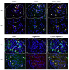
Figure 1
Figure 1
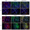
CD68/iNOS and CD68/arginase-1 co-expression in the kidneys of patients with IgAV nephritis. Immunofluorescence assay shows double labelling with anti-CD68 mouse IgG1 (red) and anti-iNOS rabbit IgG (green) in the glomeruli-G (A) and in the tubules (B) or double labelling with anti-CD68 rabbit IgG (green) and anti-arginase-1 mouse IgG3 (red) in the glomeruli (C) and in the tubules (D). White arrows indicate cells with red or green fluorescence. The co-expression of CD68 and iNOS or arginase-1 is visualised in yellow in the last column (yellow arrows). The nuclei are stained blue with 40,6-diamidino-2-phenylindole. White dashed curves mark the boundary of the glomeruli (G). One of eight experiments conducted in each group is shown. Magnification was achieved by the Olympus UPlan objective lens Apo100×/1.35 Oil Iris (×1000).
Figure 2
Figure 2
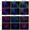
Spatial relationships among CD68, CD3, and CD56 positive cells, and CD56 and NKp44 co-expression in glomeruli (G) of patients with IgAV nephritis. Immunofluorescence shows double-labelled anti-CD68 mouse IgG1(red, white arrows) and anti-CD3 rabbit IgG (green, white arrowheads) (A), anti-CD68 mouse IgG1(red, white arrows) and anti-CD56 rabbit IgG (green, white arrowheads) (B), and anti-CD56 rabbit polyclonal (red, white arrows) and anti-NKp44 mouse IgG2a (green, white arrows) (C). The co-expression of CD56 and NKp44 is shown in yellow in the last column (yellow arrows). Nuclei are stained blue with 40,6-diamidino-2-phenylindole. White dashed curves mark the boundary of the glomeruli (G). One of the eight experiments conducted in each group is presented herein. Magnification was achieved using an Olympus Uplan objective lens Apo100×/1.35 Oil Iris (×1000).
Figure 3
Figure 3
CD56/perforin and CD3/perforin co-expression in kidney of the patients with IgAV nephritis. Immunofluorescence revealed double labelling with anti-CD56 rabbit IgG (red) and antiperforin mouse IgG2b (green) in the glomeruli (A) and tubules (B), or double labelling with anti-CD3 rabbit IgG and antiperforin mouse IgG2b in the glomeruli (C) and tubules (D). White arrows indicate cells with red or green fluorescence. Co-expression of CD56 and perforin is shown in yellow in the last column (yellow arrows). Nuclei were stained blue with 40,6-diamidino-2-phenylindole. White dashed curves mark the boundary between the glomeruli (G) and tubules (T). One of the eight experiments is shown and described. Magnification was achieved using an Olympus UPlan objective lens Apo100×/1.35 Oil Iris (×1000).
Figure 4
Figure 4
CD56/granulysin and CD3/granulysin co-expression in the kidneys of patients with IgAV nephritis. Immunofluorescence shows double labelling with anti-CD56 rabbit IgG (red) and antigranulysin mouse IgG1 (green) in the glomeruli (A) and the tubules (B), or double labelling with anti-CD3 rabbit IgG and antigranulysin mouse IgG1 in the glomeruli (C) and tubules (D). White arrows indicate cells with red or green fluorescence. Co-expression of CD56 and granulysin is shown in yellow in the last column (yellow arrows). White dashed curves mark the boundary between the glomeruli (G) and tubules (T). Nuclei were stained blue with 40,6-diamidino-2-phenylindole. One of the eight experiments conducted in each group is described above. Magnification was achieved using an Olympus UPlan objective lens Apo100×/1.35 Oil Iris (×1000).

References

    1. Jelusic M., Sestan M., Giani T., Cimaz R. New Insights and Challenges Associated With IgA Vasculitis and IgA Vasculitis With Nephritis—Is It Time to Change the Paradigm of the Most Common Systemic Vasculitis in Childhood? Front. Pediatr. 2022;10:853724. doi: 10.3389/fped.2022.853724. - DOI - PMC - PubMed
    1. Piram M., Maldini C., Biscardi S., De Suremain N., Orzechowski C., Georget E., Regnard D., Koné-Paut I., Mahr A. Incidence of IgA vasculitis in children estimated by four-source capture-recapture analysis: A population-based study. Rheumatology. 2017;56:1358–1366. doi: 10.1093/rheumatology/kex158. - DOI - PubMed
    1. Song Y., Huang X., Yu G., Qiao J., Cheng J., Wu J., Chen J. Pathogenesis of IgA Vasculitis: An Up-To-Date Review. Front. Immunol. 2021;12:771619. doi: 10.3389/fimmu.2021.771619. - DOI - PMC - PubMed
    1. Jennette J.C., Falk R.J., Bacon P.A., Basu N., Cid M.C., Ferrario F., Flores-Suarez L.F., Gross W.L., Guillevin L., Hagen E.C., et al. Overview of the 2012 revised International Chapel Hill Consensus Conference Nomenclature of Vasculitides. Arthritis Rheum. 2013;65:603–606. doi: 10.1002/art.37715. - DOI - PubMed
    1. Davin J.-C., Coppo R. Henoch–Schönlein Purpura Nephritis in Children. Nat. Rev. Nephrol. 2014;10:563–573. doi: 10.1038/nrneph.2014.126. - DOI - PubMed