. 2024 Feb 15;25(4):2313.
doi: 10.3390/ijms25042313.

Antioxidant Efficacy of Hwangryunhaedok-tang through Nrf2 and AMPK Signaling Pathway against Neurological Disorders In Vivo and In Vitro

Affiliations

Antioxidant Efficacy of Hwangryunhaedok-tang through Nrf2 and AMPK Signaling Pathway against Neurological Disorders In Vivo and In Vitro

Su-Jin Bae et al. Int J Mol Sci. .

Abstract

Alzheimer's disease (AD) is a representative cause of dementia and is caused by neuronal loss, leading to the accumulation of aberrant neuritic plaques and the formation of neurofibrillary tangles. Oxidative stress is involved in the impaired clearance of amyloid beta (Aβ), and Aβ-induced oxidative stress causes AD by inducing the formation of neurofibrillary tangles. Hwangryunhaedok-tang (HHT, Kracie K-09®), a traditional herbal medicine prescription, has shown therapeutic effects on various diseases. However, the studies of HHT as a potential treatment for AD are insufficient. Therefore, our study identified the neurological effects and mechanisms of HHT and its key bioactive compounds against Alzheimer's disease in vivo and in vitro. In a 5xFAD mouse model, our study confirmed that HHT attenuated cognitive impairments in the Morris water maze (MWM) test and passive avoidance (PA) test. In addition, the prevention of neuron impairment, reduction in the protein levels of Aβ, and inhibition of cell apoptosis were confirmed with brain tissue staining. In HT-22 cells, HHT attenuates tBHP-induced cytotoxicity, ROS generation, and mitochondrial dysfunction. It was verified that HHT exerts a neuroprotective effect by activating signaling pathways interacting with Nrf2, such as MAPK/ERK, PI3K/Akt, and LKB1/AMPK. Among the components, baicalein, a bioavailable compound of HHT, exhibited neuroprotective properties and activated the Akt, AMPK, and Nrf2/HO-1 pathways. Our findings indicate a mechanism for HHT and its major bioavailable compounds to treat and prevent AD and suggest its potential.

Keywords: AMPK; Alzheimer’s disease; Hwangryunhaedok-tang; Nrf2; baicalein.


Conflict of interest statement

The funders had no role in the design of the study; in the collection, analyses, or interpretation of data; in the writing of the manuscript; or in the decision to publish the results.

Figures

Figure 1
Figure 1
Effects of HHT, resulting in reduced cognitive impairments in 5xFAD transgenic AD mice. (A) Schematic timeline of experiment; (B) representative tracks of the test during the probe trial on day 5; (C) escape latency for 5 days; *** p < 0.001 (CTL vs. Veh), ### p < 0.001 (Veh vs. HHT 100 mg/kg), and $$ p < 0.01 and $$$ p < 0.001 (Veh vs. HHT 100 mg/kg). (D) Mice arriving at the platform in a Morris water maze (MWM) test and (E) step-through latency in passive avoidance (PA) test. Experimental data represent the means ± S.E.M. ** p < 0.01 and *** p < 0.001 (vs. CTL); # p < 0.05 and ### p < 0.001 (vs. Veh). Veh, 5xFAD mice; Done, donepezil.
Figure 2
Figure 2
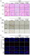
HHT decreased neuronal damage, Aβ accumulation, and the expression in hippocampus of 5xFAD mice. (A) Representative images of H–E staining in the CA1 and CA3 regions of the hippocampus among experimental groups. Images were taken through a compound light microscope using a 100× (upper) and 200× (middle and lower) objective. (B) Representative images of Aβ protein levels in the CA1 and DG hippocampal regions among experimental groups. Images were taken through a compound light microscope using a 100× (upper) and 200× (middle and lower) objective. (C) Representative immunofluorescence images for detection of apoptosis cell by TUNEL staining assay (red) and Nuclei were stained blue (DAPI) in the hippocampus. Images were taken through a compound light microscope using a 100× and 200× (DG region) objective.
Figure 3
Figure 3
Neuroprotective effect of HHT on tBHP-induced cytotoxicity, ROS generation, and mitochondrial dysfunction. (A) Cell viability was examined using the MTT assay. HT-22 cells were treated with HHT (3, 10, 30, 100, and 300 μg/mL) for 1 h, followed by incubation with 100 μm tBHP for 6 h. Data are presented as the means ± SDs of three replicate experiments (** p < 0.01, significant difference compared to control; ## p < 0.01, significant difference compared to tBHP). (B) HT-22 cells were incubated with or without 300 μg/mL HHT for 1 h, then incubated with 100 μm tBHP for 6 h. Immunoblot analysis for apoptosis-associated proteins was performed with HT-22 cell lysates. (C) Cell viability was measured using a fluorescence microscope after staining with calcein AM (0.5 μg/mL) and propidium iodide (0.5 μg/mL) for 30 min. (D) HT-22 cells were treated as described in Figure 3B. (D) Cellular ROS generation was measured using a DCFH-DA assay kit. (E,F) The fluorescence intensity of rhodamine 123 in the mitochondrial inner membrane was analyzed using a flow cytometer. Rhodamine 123 (0.05 μg/mL) staining was carried out for 1 h. Data are presented as the means ± SDs of three replicate experiments (** p < 0.01, significant difference compared to control; ## p < 0.01, significant difference compared to tBHP).
Figure 4
Figure 4
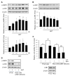
Effects of HHT on the activation of the Nrf2/HO-1 signaling pathway. The HT-22 cells were treated with (A) 30, 100, and 300 μg/mL HHT for 6 h and (B) 300 μg/mL HHT for the indicated time. Subsequently, nuclear fractionation was performed for the immunoblotting analysis of the accumulation of Nrf2 in the cell nucleus. Lamin A/C was used for the immunoblotting analysis for equal loading control of cell nucleus proteins. (C) Immunoblotting analysis of HO-1 was performed with lysates of HT-22 cells treated with 300 μg/mL HHT for 30 min to 12 h. Expression of Nrf2 and HO-1 are presented as means ± SD of three replicate experiments (* p < 0.05; ** p < 0.01, significant difference compared to control). (D) Immunoblotting analysis of Nrf2 was performed using lysates of HT-22 cells treated with 300 μg/mL HHT and 20 μM IsoLQ for 3 h each. (E) Immunoblotting analyses of p-ERK and p-Akt were performed with HT-22 cells lysates treated with 300 μg/mL HHT for 10 min to 6 h. (F) HT-22 cells were treated or not with LY294002 (30 μM), PD98059 (30 μM), and compound C (5 μM) for 1 h, followed by incubation with 300 μg/mL HHT for 1 h. Additionally, after incubation with 100 μm tBHP for 6 h, the MTT assay was performed. Data are presented as means ± SDs of three replicate experiments (** p < 0.01, significant difference compared to control; ## p < 0.01, significant difference compared to tBHP; ++ p < 0.01, significant difference compared to tBHP + HHT).
Figure 5
Figure 5
Effects of HHT on the activation of AMPK signaling pathway. Immunoblotting analyses of p-AMPK, p-ACC (A), and p-LKB1 (B) were performed using HT-22 cell lysates treated with 300 μg/mL HHT for 10 min to 6 h. Expressions of p-AMPK, p-ACC, and p-LKB1 are presented as means ± SDs of three replicate experiments (* p < 0.05; ** p < 0.01, significant difference compared to the control). (C) HT-22 and LKB1-deficient HeLa cells were incubated with 300 μg/mL HHT, followed by MTT assay. Data are presented as means ± SDs of the three replicate experiments (** p < 0.01: significant difference compared to control; ## p < 0.01: significant difference compared to tBHP). (D) Immunoblotting analysis was performed with HT-22 and HeLa cell lysates incubated with 300 μg/mL HHT for 1 h.
Figure 6
Figure 6
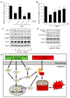
Effects of baicalein, a key compound in HHT, on neuroprotection and activation of AMPK signaling pathway. (A) HT-22 cells were treated with 10 μM each of baicalein, wogonin, and geniposide for 1 h, and then incubated with 100 μm tBHP for 6 h, followed by the MTT assay. (B) HT-22 cells were treated with baicalein (1, 3, and 10 μM) for 1 h, followed by the next sequence described in Figure 6A. Data are presented as the means ± SDs of three replicate experiments (** p < 0.01, significant difference compared to control; ## p < 0.01, significant difference compared to tBHP). (C) Immunoblotting analysis to confirm activation of the Akt and AMPK signaling pathways was performed with HT-22 cell lysates. Cells were treated with 10 μM baicalein for 10 min to 6 h. (D) HT-22 cells were treated with 10 μM baicalein for 3, 6, and 12 h, followed by immunoblotting analysis of Nrf2 in nuclear fractions and HO-1 in whole lysates, respectively. (E) Schematic diagram of neuroprotective effects of HHT.

Similar articles

Cited by

References

    1. GBD 2019 Dementia Forecasting Collaborators Estimation of the global prevalence of dementia in 2019 and forecasted prevalence in 2050: An analysis for the Global Burden of Disease Study 2019. Lancet Public Health. 2022;7:e105–e125. doi: 10.1016/S2468-2667(21)00249-8. - DOI - PMC - PubMed
    1. van der Kant R., Goldstein L.S.B., Ossenkoppele R. Amyloid-β-independent regulators of tau pathology in Alzheimer disease. Nat. Rev. Neurosci. 2020;21:21–35. doi: 10.1038/s41583-019-0240-3. - DOI - PubMed
    1. Long J.M., Holtzman D.M. Alzheimer Disease: An Update on Pathobiology and Treatment Strategies. Cell. 2019;179:312–339. doi: 10.1016/j.cell.2019.09.001. - DOI - PMC - PubMed
    1. Busche M.A., Hyman B.T. Synergy between amyloid-β and tau in Alzheimer’s disease. Nat. Neurosci. 2020;23:1183–1193. doi: 10.1038/s41593-020-0687-6. - DOI - PMC - PubMed
    1. Scheltens P., De Strooper B., Kivipelto M., Holstege H., Chételat G., Teunissen C.E., Cummings J., van der Flier W.M. Alzheimer’s disease. Lancet. 2021;397:1577–1590. doi: 10.1016/S0140-6736(20)32205-4. - DOI - PMC - PubMed

MeSH terms