

Multiple Lines of Evidence Support 199 SARS-CoV-2 Positively Selected Amino Acid Sites
- PMID: 38397104
- PMCID: PMC10889775
- DOI: 10.3390/ijms25042428
Multiple Lines of Evidence Support 199 SARS-CoV-2 Positively Selected Amino Acid Sites
Abstract
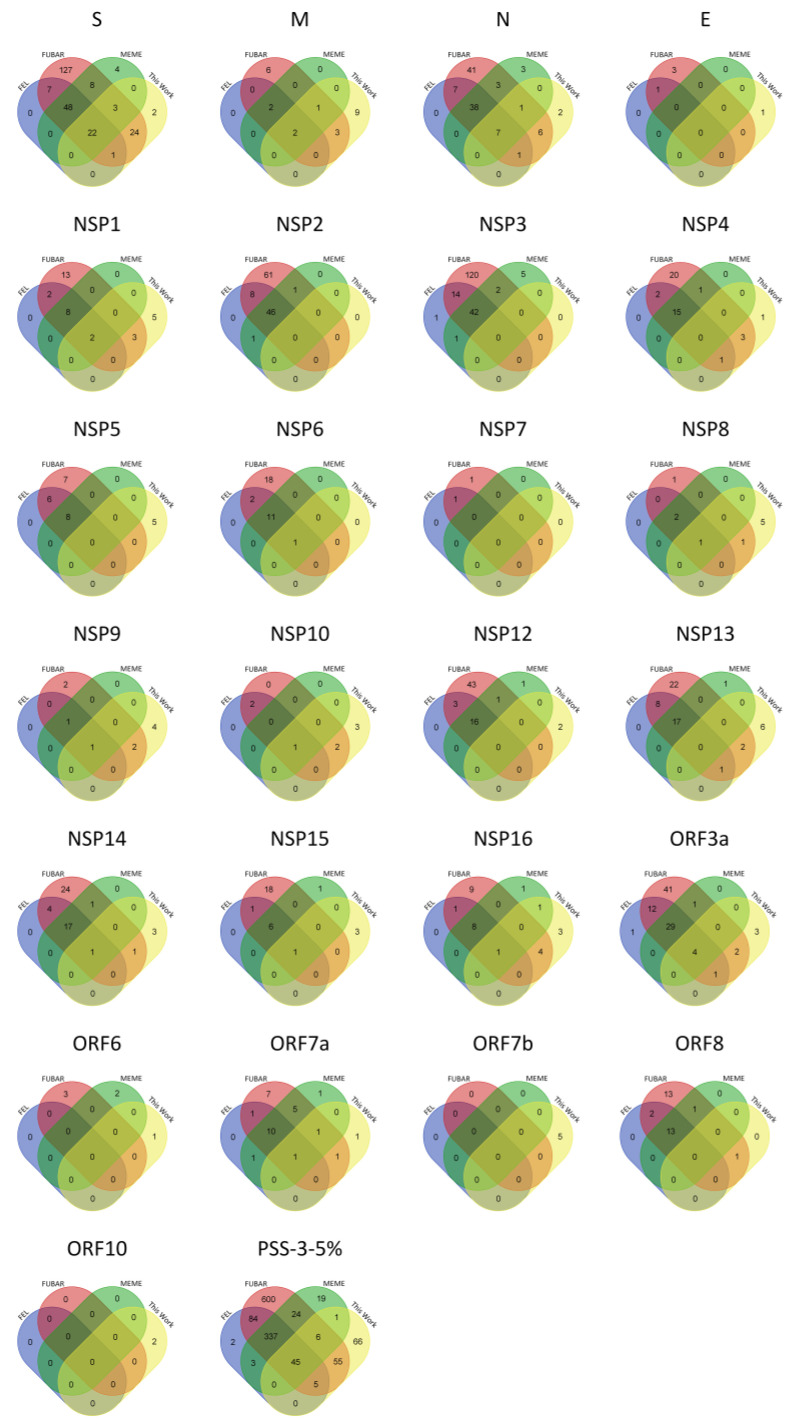
SARS-CoV-2 amino acid variants that contribute to an increased transmissibility or to host immune system escape are likely to increase in frequency due to positive selection and may be identified using different methods, such as codeML, FEL, FUBAR, and MEME. Nevertheless, when using different methods, the results do not always agree. The sampling scheme used in different studies may partially explain the differences that are found, but there is also the possibility that some of the identified positively selected amino acid sites are false positives. This is especially important in the context of very large-scale projects where hundreds of analyses have been performed for the same protein-coding gene. To account for these issues, in this work, we have identified positively selected amino acid sites in SARS-CoV-2 and 15 other coronavirus species, using both codeML and FUBAR, and compared the location of such sites in the different species. Moreover, we also compared our results to those that are available in the COV2Var database and the frequency of the 10 most frequent variants and predicted protein location to identify those sites that are supported by multiple lines of evidence. Amino acid changes observed at these sites should always be of concern. The information reported for SARS-CoV-2 can also be used to identify variants of concern in other coronaviruses.
Keywords: SARS-CoV-2; coronaviruses; positively selected amino acid sites.
Conflict of interest statement
The authors declare no conflicts of interest. The funders had no role in the design of the study; in the collection, analyses, or interpretation of data; in the writing of the manuscript; or in the decision to publish the results.
Figures
Similar articles
-
Dissecting Naturally Arising Amino Acid Substitutions at Position L452 of SARS-CoV-2 Spike.J Virol. 2022 Oct 26;96(20):e0116222. doi: 10.1128/jvi.01162-22. Epub 2022 Oct 10. J Virol. 2022. PMID: 36214577 Free PMC article.
-
COV2Var, a function annotation database of SARS-CoV-2 genetic variation.Nucleic Acids Res. 2024 Jan 5;52(D1):D701-D713. doi: 10.1093/nar/gkad958. Nucleic Acids Res. 2024. PMID: 37897356 Free PMC article.
-
In Silico Analysis of the Effects of Omicron Spike Amino Acid Changes on the Interactions with Human Proteins.Molecules. 2022 Jul 28;27(15):4827. doi: 10.3390/molecules27154827. Molecules. 2022. PMID: 35956778 Free PMC article.
-
Understanding the Secret of SARS-CoV-2 Variants of Concern/Interest and Immune Escape.Front Immunol. 2021 Nov 5;12:744242. doi: 10.3389/fimmu.2021.744242. eCollection 2021. Front Immunol. 2021. PMID: 34804024 Free PMC article.
-
Evolution of SARS-CoV-2 Variants: Implications on Immune Escape, Vaccination, Therapeutic and Diagnostic Strategies.Viruses. 2023 Apr 10;15(4):944. doi: 10.3390/v15040944. Viruses. 2023. PMID: 37112923 Free PMC article. Review.
Cited by
-
Patterns of Recombination in Coronaviruses.Int J Mol Sci. 2025 Jun 11;26(12):5595. doi: 10.3390/ijms26125595. Int J Mol Sci. 2025. PMID: 40565057 Free PMC article.
References
-
- Gorbalenya A.E., Baker S.C., Baric R.S., de Groot R.J., Drosten C., Gulyaeva A.A., Haagmans B.L., Lauber C., Leontovich A.M., Neuman B.W., et al. The species Severe acute respiratory syndrome-related coronavirus: Classifying 2019-nCoV and naming it SARS-CoV-2. Nat. Microbiol. 2020;5:536–544. doi: 10.1038/s41564-020-0695-z. - DOI - PMC - PubMed
MeSH terms
Substances
Supplementary concepts
Grants and funding
LinkOut - more resources
Full Text Sources
Medical
Miscellaneous
