

Myconoside and Calceolarioside E Restrain UV-Induced Skin Photoaging by Activating NRF2-Mediated Defense Mechanisms
- PMID: 38397118
- PMCID: PMC10888667
- DOI: 10.3390/ijms25042441
Myconoside and Calceolarioside E Restrain UV-Induced Skin Photoaging by Activating NRF2-Mediated Defense Mechanisms
Abstract
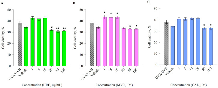
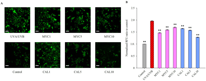
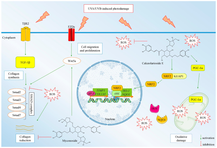
Chronic and excessive ultraviolet (UVA/UVB) irradiation exposure is known as a major contributor to premature skin aging, which leads to excessive reactive oxygen species generation, disturbed extracellular matrix homeostasis, DNA damage, and chronic inflammation. Sunscreen products are the major preventive option against UVR-induced photodamage, mostly counteracting the acute skin effects and only mildly counteracting accelerated aging. Therefore, novel anti-photoaging and photopreventive compounds are a subject of increased scientific interest. Our previous investigations revealed that the endemic plant Haberlea rhodopensis Friv. (HRE) activates the antioxidant defense through an NRF2-mediated mechanism in neutrophiles. In the present study, we aimed to investigate the photoprotective potential of HRE and two of its specialized compounds-the phenylethanoid glycosides myconoside (MYC) and calceolarioside E (CAL)-in UVA/UVB-stimulated human keratinocytes in an in vitro model of photoaging. The obtained data demonstrated that the application of HRE, MYC, and CAL significantly reduced intracellular ROS formation in UVR-exposed HaCaT cells. The NRF2/PGC-1α and TGF-1β/Smad/Wnt signaling pathways were pointed out as having a critical role in the observed CAL- and MYC-induced photoprotective effect. Collectively, CAL is worth further evaluation as a potent natural NRF2 activator and a promising photoprotective agent that leads to the prevention of UVA/UVB-induced premature skin aging.
Keywords: NRF2; calceolarioside; keratinocytes; myconoside; photoaging; ultraviolet radiation.
Conflict of interest statement
The authors declare no conflicts of interest.
Figures


Similar articles
-
Biotechnologically-Produced Myconoside and Calceolarioside E Induce Nrf2 Expression in Neutrophils.Int J Mol Sci. 2021 Feb 10;22(4):1759. doi: 10.3390/ijms22041759. Int J Mol Sci. 2021. PMID: 33578811 Free PMC article.
-
Limonene protects human skin keratinocytes against UVB-induced photodamage and photoaging by activating the Nrf2-dependent antioxidant defense system.Environ Toxicol. 2022 Dec;37(12):2897-2909. doi: 10.1002/tox.23646. Epub 2022 Sep 5. Environ Toxicol. 2022. PMID: 36063024
-
Anti-Photoaging Effect of Phaseolus angularis L. Extract on UVB-Exposed HaCaT Keratinocytes and Possibilities as Cosmetic Materials.Molecules. 2023 Feb 1;28(3):1407. doi: 10.3390/molecules28031407. Molecules. 2023. PMID: 36771069 Free PMC article.
-
Effect of ultraviolet radiation on the Nrf2 signaling pathway in skin cells.Int J Radiat Biol. 2021;97(10):1383-1403. doi: 10.1080/09553002.2021.1962566. Epub 2021 Aug 19. Int J Radiat Biol. 2021. PMID: 34338112 Review.
-
Roles of the KEAP1-NRF2 system in mammalian skin exposed to UV radiation.Toxicol Appl Pharmacol. 2018 Dec 1;360:69-77. doi: 10.1016/j.taap.2018.09.038. Epub 2018 Sep 28. Toxicol Appl Pharmacol. 2018. PMID: 30268578 Review.
Cited by
-
Vorinostat attenuates UVB-induced skin senescence by modulating NF-κB and mTOR signaling pathways.Sci Rep. 2025 Mar 29;15(1):10905. doi: 10.1038/s41598-025-95624-4. Sci Rep. 2025. PMID: 40158057 Free PMC article.
-
Atractylodin mitigates UVB radiation-induced oxidative stress and photoaging responses by enhancing NrF2 signaling in human epidermal keratinocytes.Arch Dermatol Res. 2024 Dec 30;317(1):160. doi: 10.1007/s00403-024-03657-y. Arch Dermatol Res. 2024. PMID: 39738877
-
The total extract of Abelmoschus manihot (L.) medic flowers (TEA) mediated Nrf2-TFAM signalling to regulate mitochondrial antioxidant mechanism.Sci Rep. 2025 Jan 10;15(1):1614. doi: 10.1038/s41598-024-84022-x. Sci Rep. 2025. PMID: 39794424 Free PMC article.
References
-
- de Assis L., Tonolli P., Moraes M., Baptista M., Castrucci A. How does the skin sense sun light? An integrative view of lightsensing molecules. J. Photochem. Photobiol. C-Photochem. Rev. 2021;47:100403. doi: 10.1016/j.jphotochemrev.2021.100403. - DOI
-
- Kahremany S., Hofmann L., Gruzman A., Dinkova-Kostova A.T., Cohen G. NRF2 in dermatological disorders: Pharmacological activation for protection against cutaneous photodamage and photodermatosis. Free. Radic. Biol. Med. 2022;188:262–276. doi: 10.1016/j.freeradbiomed.2022.06.238. - DOI - PMC - PubMed
MeSH terms
Substances
Grants and funding
LinkOut - more resources
Full Text Sources
Medical
