. 2024 Jan 29;15(2):183.
doi: 10.3390/genes15020183.

The Impact of Psilocybin on High Glucose/Lipid-Induced Changes in INS-1 Cell Viability and Dedifferentiation

Affiliations

The Impact of Psilocybin on High Glucose/Lipid-Induced Changes in INS-1 Cell Viability and Dedifferentiation

Esmaeel Ghasemi Gojani et al. Genes (Basel). .

Abstract

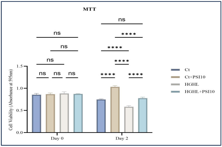
Serotonin emerges as a pivotal factor influencing the growth and functionality of β-cells. Psilocybin, a natural compound derived from mushrooms of the Psilocybe genus, exerts agonistic effects on the serotonin 5-HT2A and 5-HT2B receptors, thereby mimicking serotonin's behavior. This study investigates the potential impacts of psilocybin on β-cell viability, dedifferentiation, and function using an in vitro system. The INS-1 832/13 Rat Insulinoma cell line underwent psilocybin pretreatment, followed by exposure to high glucose-high lipid (HG-HL) conditions for specific time periods. After being harvested from treated cells, total transcript and cellular protein were utilized for further investigation. Our findings implied that psilocybin administration effectively mitigates HG-HL-stimulated β-cell loss, potentially mediated through the modulation of apoptotic biomarkers, which is possibly related to the mitigation of TXNIP, STAT-1, and STAT-3 phosphorylation. Furthermore, psilocybin exhibits the capacity to modulate the expression of key genes associated with β-cell dedifferentiation, including Pou5f1 and Nanog, indicating its potential in attenuating β-cell dedifferentiation. This research lays the groundwork for further exploration into the therapeutic potential of psilocybin in Type II diabetes intervention.

Keywords: diabetes; psilocybin; β-cell dedifferentiation; β-cell loss.


Conflict of interest statement

The authors declare no conflicts of interest. The funders had no role in the design of the study; in the collection, analyses, or interpretation of data; in the writing of the manuscript; or in the decision to publish the results.

Figures

Figure 1
Figure 1
The impact of 48 h treatment with 10 μM psilocybin on the survival of β-cells challenged by HG-HL. INS-1 cells were pretreated with 10 μM psilocybin, followed by induction with HG-HL (400 μM PA + 25 mM glucose) for the next 48 h. Afterward, the cells were incubated with the MTT reagent for 4 h, followed by the incubation with MTT solubilizing buffer overnight. The results are depicted as the mean value with standard deviation, based on three experiments (N = 3). Abbreviations used are Ct (control), PSI (psilocybin), and HG-HL (high glucose + high lipid). Significance is denoted by asterisks, with four indicating p < 0.0001, while “ns” denotes non-significance.
Figure 2
Figure 2
The immunoblot analysis of apoptotic biomarkers in HG-HL-challenged β-cells in response to psilocybin. (a) INS-1 cells were exposed to 10 μM psilocybin, followed by incubation with HG-HL for the next 48 h. Subsequently, the treated cells were used for protein extraction for Western blot analysis. The response of Pro-Caspase-9, C-Caspase-9, Pro-Caspase-7, C-Caspase-7, C-Capse-3, C-PARP, Bcl-2, Bim and Bax to 10 μM psilocybin in HG-HL-challenged β-cells, with normalization to housekeeping proteins. (b) Depicted are Western blot images of the apoptotic biomarkers. (c) Caspase 3/7 activity assay performed on β-cells challenged by HG-HL and treated with 10 μM psilocybin showed the mitigative impact of psilocybin on the elevated Caspase-3 and Caspase-7 activity in HG-HL-challenged β-cells. The results are depicted as the mean value with standard deviation, based on three measurements (N = 3). Abbreviations used are Ct (control), PSI (psilocybin), and HG-HL (high glucose + high lipid). Significance is denoted by asterisks, with two implying p < 0.01 and four indicating p < 0.0001.
Figure 3
Figure 3
The immunoblot analysis of P-STAT-3, T-STAT-3, and P-STAT-1 in HG-HL- β-cells in response to 10 μM psilocybin. The administration of 10 μM psilocybin resulted in significant reduction in the levels of P-STAT-3, T-STAT-3, and P-STAT-1 in HG-HL-challenged β-cells. Following band quantification using ImageJ, the obtained results were normalized to housekeeping proteins, including α-Tubulin and β-Actin. The results are depicted as the mean value with standard deviation, based on three measurements (N = 3). Abbreviation: Ct (control), PSI (psilocybin) and HG-HL (high glucose + high lipid). Significance is denoted by asterisks, with four implying p < 0.0001.
Figure 4
Figure 4
The impact of 10 μM psilocybin on GSIS of HG-HL-challenged β-cells. GSIS response of HG-HL-induced β-cell to psilocybin in media comprising 2.5 mM glucose and 16.5 mM glucose. Psilocybin did not exhibit any positive effects on the impaired GSIS in HG-HL-stimulated β-cells. The results are depicted as the mean value with standard deviation, based on three measurements (N = 3). Abbreviations used are Ct (control), PSI (psilocybin), and HG-HL (high glucose + high lipid). Significance is denoted by asterisks, with three implying p < 0.001 and four indicating p < 0.0001, while “ns” denotes non-significance.
Figure 5
Figure 5
The alterations of P-FOXO1, FOXO1, TXNIP, and PDX-1 levels in HG-HL-stimulated β-cell in response to psilocybin. The administration of 10 μM psilocybin resulted in the reduction in P-FOXO1, FOXO1, TXNIP, and PDX-1 levels in HG-HL-induced β-cells. The findings are illustrated as the average value with standard deviation, derived from three measurements (N = 3). Abbreviations include Ct (control), PSI (psilocybin), and HG-HL (high glucose + high lipid). Significance is indicated by asterisks, where two asterisks indicate p < 0.01, three indicate p < 0.001, and four indicate p < 0.0001.
Figure 6
Figure 6
The qPCR analysis assessed the expression of key dedifferentiation-associated transcripts in both HG-HL-stimulated and unstimulated β-cells following treatment with 10 µM psilocybin. INS-1 β-cells underwent a 2 h pre-treatment with psilocybin, followed by a 24 h incubation in HG-HL conditions (250 μM palmitic acid + 25 mM glucose). Total RNA was extracted from both treated and untreated cells and utilized for qRT-PCR. The figure illustrates the normalized mRNA expression levels of Ins1, Ins2, PDX-1, FOXO1, NEUROD1, MafA, Slc2A2, Nanog, and Pou5f1 in both HG-HL-stimulated and unstimulated β-cells treated with 10 µM psilocybin. The results are shown as the average value with the standard deviation, from three replicates (N = 3). Abbreviations include Ct (control), PSI (psilocybin), and HG-HL (high glucose + high lipid). Significance is marked by asterisks: one for p < 0.05, two for p < 0.01, three for p < 0.001, four for p < 0.0001, and “ns” for non-significant outcomes.

References

    1. Holt R.I., Cockram C., Flyvbjerg A., Goldstein B.J. Textbook of Diabetes. John Wiley & Sons; Hoboken, NJ, USA: 2017.
    1. Back S.H., Kaufman R.J. Endoplasmic reticulum stress and type 2 diabetes. Annu. Rev. Biochem. 2012;81:767–793. doi: 10.1146/annurev-biochem-072909-095555. - DOI - PMC - PubMed
    1. Ghasemi-Gojani E., Kovalchuk I., Kovalchuk O. Cannabinoids and terpenes for diabetes mellitus and its complications: From mechanisms to new therapies. Trends Endocrinol. Metab. 2022;33:828–849. doi: 10.1016/j.tem.2022.08.003. - DOI - PubMed
    1. Bensellam M., Jonas J.-C., Laybutt D.R. Mechanisms of β-cell dedifferentiation in diabetes: Recent findings and future research directions. J. Endocrinol. 2018;236:R109–R143. doi: 10.1530/JOE-17-0516. - DOI - PubMed
    1. Amo-Shiinoki K., Tanabe K., Hoshii Y., Matsui H., Harano R., Fukuda T., Takeuchi T., Bouchi R., Takagi T., Hatanaka M. Islet cell dedifferentiation is a pathologic mechanism of long-standing progression of type 2 diabetes. JCI Insight. 2021;6:e143791. doi: 10.1172/jci.insight.143791. - DOI - PMC - PubMed

Publication types

Grants and funding