

Dilated Cardiomyopathy Due to Alimentary Iron Deficiency
- PMID: 38397308
- PMCID: PMC10887770
- DOI: 10.3390/children11020196
Dilated Cardiomyopathy Due to Alimentary Iron Deficiency
Abstract
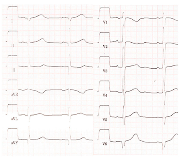
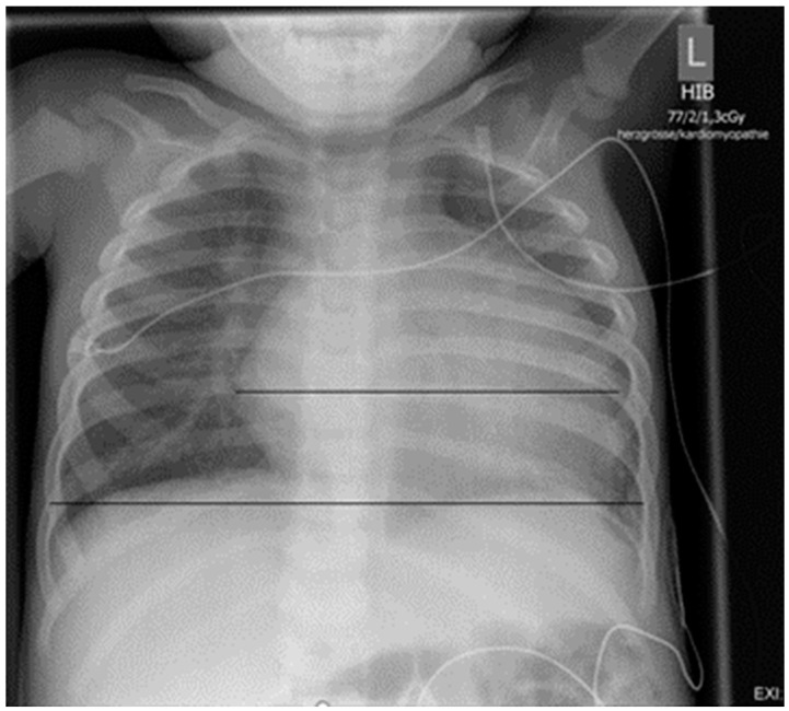
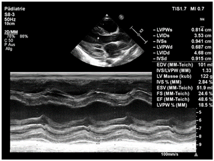
Dilated cardiomyopathy (DCM) is a severe condition, characterised by left ventricular dilation and systolic dysfunction, necessitating heart transplantation when all other treatment options fail. This case report describes a 2-year-old girl initially presenting with oedema, listlessness, and severe iron deficiency anaemia. She was diagnosed with DCM. Extensive diagnostic workup ruled out other causes, leading to the suspicion of DCM due to alimentary iron deficiency. This was confirmed by the parents' report that the girl was fed almost exclusively with low-fat cow's milk. Prompt treatment, including packed red cell transfusion, iron supplementation, and heart failure medications (diuretics, ACE inhibitors, beta blockers, and aldosterone antagonists), resulted in significant improvement in cardiac function within days. This report demonstrates the potential risks of alimentary iron deficiency, the most common cause of microcytic hypochromic anaemia in young children, which might even result in the development of life-threatening cardiac dysfunction in extreme cases.
Keywords: anaemia; cardiomyopathy; iron; nutrition; paediatric cardiology.
Conflict of interest statement
The authors declare no conflicts of interest.
Figures
References
-
- Lipshultz S.E., Law Y.M., Asante-Korang A., Austin E.D., Dipchand A.I., Everitt M.D., Hsu D.T., Lin K.-Y., Price J.F., Wilkinson J.D., et al. Cardiomyopathy in Children. Classification and Diagnosis. A Scientific Statement from the American Heart Association. Circulation. 2019;140:e9–e68. doi: 10.1161/CIR.0000000000000682. - DOI - PubMed
-
- Koletzko B., Speer C.P., Gahr M., Dötsch J. Grundlagen der Ernährung. In: Speer C.P., Gahr M., Dötsch J., editors. Pädiatrie. Springer; Berlin/Heidelberg, Germany: 2019. pp. 25–39.
-
- World Health Organisation (WHO) [(accessed on 2 December 2023)]. Available online: https://www.who.int/news-room/fact-sheets/detail/infant-and-young-child-....
Publication types
LinkOut - more resources
Full Text Sources
Miscellaneous
