Review
. 2024 Jan 24;14(2):145.
doi: 10.3390/biom14020145.

Unveiling the Protective Role of Melatonin in Osteosarcoma: Current Knowledge and Limitations

Affiliations
Review

Unveiling the Protective Role of Melatonin in Osteosarcoma: Current Knowledge and Limitations

Nojoud Al-Ansari et al. Biomolecules. .

Abstract

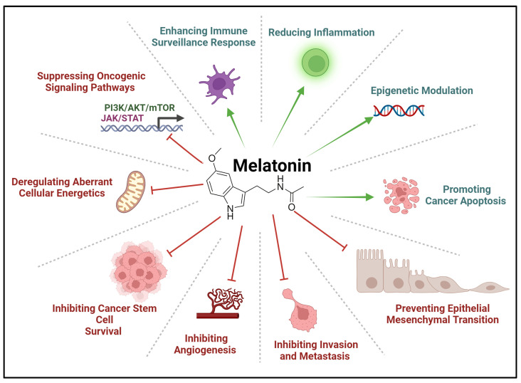
Melatonin, an endogenous neurohormone produced by the pineal gland, has received increased interest due to its potential anti-cancer properties. Apart from its well-known role in the sleep-wake cycle, extensive scientific evidence has shown its role in various physiological and pathological processes, such as inflammation. Additionally, melatonin has demonstrated promising potential as an anti-cancer agent as its function includes inhibition of tumorigenesis, induction of apoptosis, and regulation of anti-tumor immune response. Although a precise pathophysiological mechanism is yet to be established, several pathways related to the regulation of cell cycle progression, DNA repair mechanisms, and antioxidant activity have been implicated in the anti-neoplastic potential of melatonin. In the current manuscript, we focus on the potential anti-cancer properties of melatonin and its use in treating and managing pediatric osteosarcoma. This aggressive bone tumor primarily affects children and adolescents and is treated mainly by surgical and radio-oncological interventions, which has improved survival rates among affected individuals. Significant disadvantages to these interventions include disease recurrence, therapy-related toxicity, and severe/debilitating side effects that the patients have to endure, significantly affecting their quality of life. Melatonin has therapeutic effects when used for treating osteosarcoma, attributed to its ability to halt cancer cell proliferation and trigger apoptotic cell death, thereby enhancing chemotherapeutic efficacy. Furthermore, the antioxidative function of melatonin alleviates harmful side effects of chemotherapy-induced oxidative damage, aiding in decreasing therapeutic toxicities. The review concisely explains the many mechanisms by which melatonin targets osteosarcoma, as evidenced by significant results from several in vitro and animal models. Nevertheless, if further explored, human trials remain a challenge that could shed light and support its utility as an adjunctive therapeutic modality for treating osteosarcoma.

Keywords: anti-cancer therapeutics; apoptosis; bone cancer; melatonin; osteosarcoma.


Conflict of interest statement

The authors declare no conflicts of interest. The funders had no role in the design of the study; in the collection, analyses, or interpretation of data; in the writing of the manuscript, or in the decision to publish the article.

Figures

Figure 1
Figure 1
Mechanism of action of melatonin. The image was adapted and modified from [12,19,20]. Melatonin enters the cell through various receptors on cellular surfaces (such as MT1/2 and GLUT1 or passively diffuses into the cells and organelles. Melatonin utilizes receptors such as MT1/MT2, cytoplasmic receptor quinone reductase II, and nuclear receptor RORa/RZR, leading to various biological effects. It also contributes to the function and regulation of processes of other organelles, such as mitochondria, exosomes, and ER. AKT = protein kinase B, cGMP = guanosine 3’,5’-cyclic monophosphate, CREB = cAMP-response element binding protein, IP3 = inositol trisphosphate, MT1 = melatonin receptor 1, MT2 = melatonin receptor 2, OXPHOS = oxidative phosphorylation. PDK = pyruvate dehydrogenase kinase, PI3K = phosphoinositide 3 kinase, PKC = protein kinase C, PKG = protein kinase G, SIRT3 = sirtuin 3, TCA = tricarboxylic acid cycle, and SOD2 = superoxide dismutase 2. The dotted arrows indicate possible transmembrane translocation of melatonin molecules while the regular arrows indicate pathway activation and progression via pathway related molecules. The ‘closed’ lines indicate pathway inhibition. Created with BioRender.com.
Figure 3
Figure 3
Signaling cascades involved in OS pathogenesis commonly such as the PI3K/Akt/mTOR, MAPK/ERK, TGFβ, Notch, Hedgehog, and NF-κB pathways have been evident in the different aspects of OS pathogenesis, summarized in the figure. These pathways can, independently or through cross-communication, aid osteosarcoma proliferation, survival, angiogenesis, migration, and invasion. Akt = protein kinase B, APC = adenomatous polyposis coli protein, Bcl2 = B-cell lymphoma 2, Bcl-xL = B-cell lymphoma-extra-large, CK1 = casein kinase 1, c-Myc = cellular myc, Co-F = co-factor, Dvl = dishevelled protein, EMT = epithelial mesenchymal transition, EGF = epidermal growth factor, ERK = extracellular signal-regulated kinase, FGF = fibroblast growth factor, GSK-3β = glycogen synthase kinase 3β, JNK = Jun N-terminal kinase, Kif = kinesin family member, MMP = matrix metallopeptidase, mTOR = mechanistic (formerly “mammalian”), NCID = NOTCH intracellular domain, NF-κB = nuclear factor kappa B, PI3K = phosphoinositide 3 kinase, R-SMAD = receptor-regulated SMAD, STAT = signal transducers and activators of transcription, target of rapamycin, SMAD = suppressor of mothers against decapentaplegic, SUFU = suppressor of fused homolog, TCF/LEF = T-cell factor/lymphoid enhancer factor, TGFβ = transforming growth factor-beta, TF = transcription factor, TSC 1/2 = tuberous sclerosis 1/2, VEGF = vascular endothelial growth factor, and WNT = wingless/integrated. Created with BioRender.com.
Figure 2
Figure 2
Biological functions of melatonin in various physiological sub-categories summarized in the illustration provided. ATP = adenosine triphosphate and ROS = reactive oxygen species. Created with BioRender.com.
Figure 4
Figure 4
Simplified summary of the pathogenesis of OS. CTGF = connective tissue growth factor, ECV = extracellular vesicles, HIF-1α = hypoxia-inducible factor 1 subunit alpha, IGF = insulin growth factor, MMP-9 = matrix metallopeptidase-9, PEDF = pigment epithelium-derived factor, PTH = parathyroid hormone, PTH-rp = parathyroid-related peptide, RANKL = receptor activator of nuclear factor kappa beta, TGF = transforming growth factor, TGFβ = transforming growth factor-beta, VEGF = vascular endothelial growth factor. Created with BioRender.com.
Figure 5
Figure 5
Effects of melatonin in cancer. These various oncostatic effects are found through preclinical and clinical studies in different cancers. Created with BioRender.com.
Figure 6
Figure 6
Oncostatic effects of melatonin in osteosarcoma. An illustrative summary of inhibitory (red) and enhanced (green) effects of melatonin. These include inhibiting cell cycle progression signaling pathways involved in OS tumorigenesis, such as SIRT, JAK-STAT, Rho/ROCK, ERK1/2, JNK, NOTCH, and Wnt-catenin. Melatonin also induces apoptosis through interactions with Fas/Fas-ligand, modifies cancer metabolism and immune response to malignancy, and modifies inflammatory conditions of the surrounding microenvironment by reducing ROS and inflammation. Finally, it enhances the sensitivity of tumors to current chemotherapies. CIC = capicua transcriptional repressor, EMT = epithelial-mesenchymal transition, ERK1/2 = extracellular signal-regulated kinase, NF-κB = nuclear factor kappa B, Rh0/ROCK = Rho-associated protein kinase, ROS = reactive oxygen species, and VEGF = vascular endothelial growth factor. Created with BioRender.com.

Similar articles

Cited by

References

    1. Arendt J., Aulinas A. Physiology of the Pineal Gland and Melatonin. In: Feingold K.R., Anawalt B., Blackman M.R., Boyce A., Chrousos G., Corpas E., de Herder W.W., Dhatariya K., Dungan K., Hofland J., et al., editors. Endotext. MDText.com, Inc.; South Dartmouth, MA, USA: 2000. - PubMed
    1. Claustrat B., Brun J., Chazot G. The basic physiology and pathophysiology of melatonin. Sleep Med. Rev. 2005;9:11–24. doi: 10.1016/j.smrv.2004.08.001. - DOI - PubMed
    1. Lu K.H., Lin R.C., Yang J.S., Yang W.E., Reiter R.J., Yang S.F. Molecular and Cellular Mechanisms of Melatonin in Osteosarcoma. Cells. 2019;8:1618. doi: 10.3390/cells8121618. - DOI - PMC - PubMed
    1. Lu X., Yu S., Chen G., Zheng W., Peng J., Huang X., Chen L. Insight into the roles of melatonin in bone tissue and bone-related diseases (Review) Int. J. Mol. Med. 2021;47:82. doi: 10.3892/ijmm.2021.4915. - DOI - PMC - PubMed
    1. Waldhauser F., Boepple P.A., Schemper M., Mansfield M.J., Crowley W.F., Jr. Serum melatonin in central precocious puberty is lower than in age-matched prepubertal children. J. Clin. Endocrinol. Metab. 1991;73:793–796. doi: 10.1210/jcem-73-4-793. - DOI - PubMed