. 2024 Feb 18;14(2):240.
doi: 10.3390/biom14020240.

Lactobacillus crispatus CCFM1339 Inhibits Vaginal Epithelial Barrier Injury Induced by Gardnerella vaginalis in Mice

Affiliations

Lactobacillus crispatus CCFM1339 Inhibits Vaginal Epithelial Barrier Injury Induced by Gardnerella vaginalis in Mice

Xiaoyan Huang et al. Biomolecules. .

Abstract

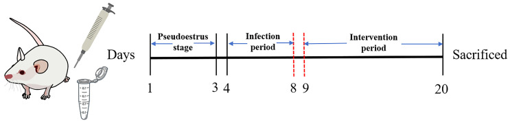
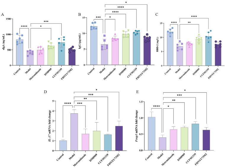
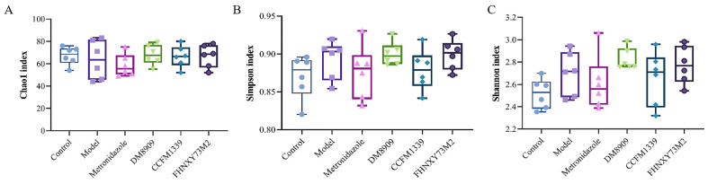
The vaginal epithelial barrier, which integrates mechanical, immune, chemical, and microbial defenses, is pivotal in safeguarding against external pathogens and upholding the vaginal microecological equilibrium. Although the widely used metronidazole effectively curtails Gardnerella vaginalis, a key pathogen in bacterial vaginosis, it falls short in restoring the vaginal barrier or reducing recurrence rates. Our prior research highlighted Lactobacillus crispatus CCFM1339, a vaginally derived Lactobacillus strain, for its capacity to modulate the vaginal epithelial barrier. In cellular models, L. crispatus CCFM1339 fortified the integrity of the cellular monolayer, augmented cellular migration, and facilitated repair. Remarkably, in animal models, L. crispatus CCFM1339 substantially abated the secretion of the barrier disruption biomarker E-cadherin (from 101.45 to 82.90 pg/mL) and increased the anti-inflammatory cytokine IL-10 (35.18% vs. the model), consequently mitigating vaginal inflammation in mice. Immunological assays in vaginal tissues elucidated increased secretory IgA levels (from 405.56 to 740.62 ng/mL) and curtailed IL-17 gene expression. Moreover, L. crispatus CCFM1339 enhanced Lactobacilli abundance and attenuated Enterobacterium and Enterococcus within the vaginal microbiome, underscoring its potential in probiotic applications for vaginal barrier regulation.

Keywords: BV; Gardnerella vaginalis; Lactobacillus crispatus; vagina epithelial barrier.


Conflict of interest statement

The authors declare that the research was conducted in the absence of any commercial or financial relationships that could be construed as a potential conflict of interest.

Figures

Figure 1
Figure 1
Animal experimental design time flow diagram.
Figure 2
Figure 2
The regulation of VK2/E6E7 monolayer barrier cells by Lactobacillus strains. (A) The Transwell model of the vaginal epithelium; (B) transepithelial electrical resistance; (C) the permeability of FD-4; (D) the wound area change treated at 24 h after the scratch; ** p < 0.01, *** p < 0.001, **** p < 0.0001 vs. model group.
Figure 3
Figure 3
Mechanical protein expression in vaginal tissue: (A) sECAD; (B) ZO-1; (C) CLDN1; (D) OCLN; * p < 0.05, ** p < 0.01, *** p < 0.001, **** p < 0.0001 vs. model group.
Figure 4
Figure 4
Changes in inflammatory factors in vaginal tissue. (A) IL-1β; (B) TNF-α; (C) MPO; (D) IL-10; * p < 0.05, ** p < 0.01, *** p < 0.001, **** p < 0.0001 vs. model group.
Figure 5
Figure 5
Effects of L. crispatus CCFM1339 on the vaginal immune barrier indicators. (A) sIgA; (B) IgG; (C) HBD-2; (D) IL-17 gene; (E) Foxp3 gene; * p < 0.05, ** p < 0.01, *** p < 0.001, **** p < 0.0001 vs. model group.
Figure 6
Figure 6
Pathological status of vaginal tissue. (A) Control; (B) model; (C) metronidazole; (D) DM8909; (E) CCFM1339; (F) FHNXY73M2; black arrow—smooth vaginal epithelium; blue arrow—local inflammatory cell infiltration of vaginal mucosa; red arrow—superficial erosion and holes.
Figure 7
Figure 7
Analysis of alpha diversity. (A) Chao1 index; (B) Simpson index; (C) Shannon index.
Figure 8
Figure 8
Analysis of beta diversity. (A) Control vs. model; (B) metronidazole vs. model; (C) DM8909 vs. model; (D) CCFM1339 vs. model; (E) FHNXY73M2 vs. model.
Figure 9
Figure 9
Analysis of different bacterial genera of vaginal microbiota. (A) Relative abundance of the vaginal microbiota at phylum level; (B) relative abundance of the vaginal microbiota at genus level; (C) branching map of species evolution; (D) histogram of LDA value distribution.

Similar articles

Cited by

References

    1. Farage M., Maibach H. Lifetime changes in the vulva and vagina. Arch. Gynecol. Obstet. 2006;273:195–202. doi: 10.1007/s00404-005-0079-x. - DOI - PubMed
    1. Ravel J., Gajer P., Abdo Z., Schneider G.M., Koenig S.S., McCulle S.L., Karlebach S., Gorle R., Russell J., Tacket C.O., et al. Vaginal microbiome of reproductive-age women. Proc. Natl. Acad. Sci. USA. 2011;108((Suppl. S1)):4680–4687. doi: 10.1073/pnas.1002611107. - DOI - PMC - PubMed
    1. Andersch-Björkman Y., Thomsson K.A., Larsson J.M.H., Ekerhovd E., Hansson G.C. Large scale identification of proteins, mucins, and their o-glycosylation in the endocervical mucus during the menstrual cycle. Mol. Cell. Proteom. 2007;6:708–716. doi: 10.1074/mcp.M600439-MCP200. - DOI - PubMed
    1. Radtke A.L., Quayle A.J., Herbst-Kralovetz M.M. Microbial products alter the expression of membrane-associated mucin and antimicrobial peptides in a three-dimensional human endocervical epithelial cell model. Biol. Reprod. 2012;87:132–141. doi: 10.1095/biolreprod.112.103366. - DOI - PMC - PubMed
    1. Gipson I.K., Moccia R., Spurr-Michaud S., Argueso P., Gargiulo A.R., Offner G.D., Keutmann H.T. The amount of muc5b mucin in cervical mucus peaks at midcycle. J. Clin. Endocrinol. Metab. 2001;86:594–600. doi: 10.1210/jc.86.2.594. - DOI - PubMed

LinkOut - more resources