. 2024 Jan 23;13(2):137.
doi: 10.3390/antiox13020137.

Liposomal Glutathione Augments Immune Defenses against Respiratory Syncytial Virus in Neonatal Mice Exposed in Utero to Ethanol

Affiliations

Liposomal Glutathione Augments Immune Defenses against Respiratory Syncytial Virus in Neonatal Mice Exposed in Utero to Ethanol

Theresa W Gauthier et al. Antioxidants (Basel). .

Abstract

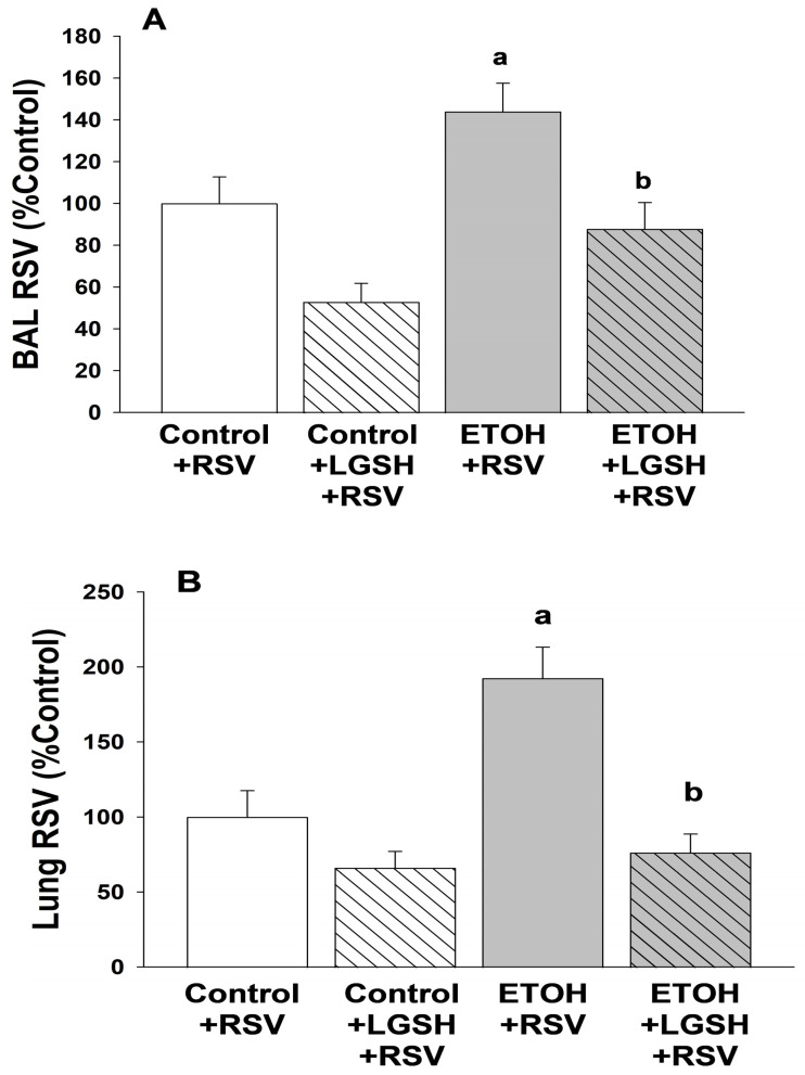
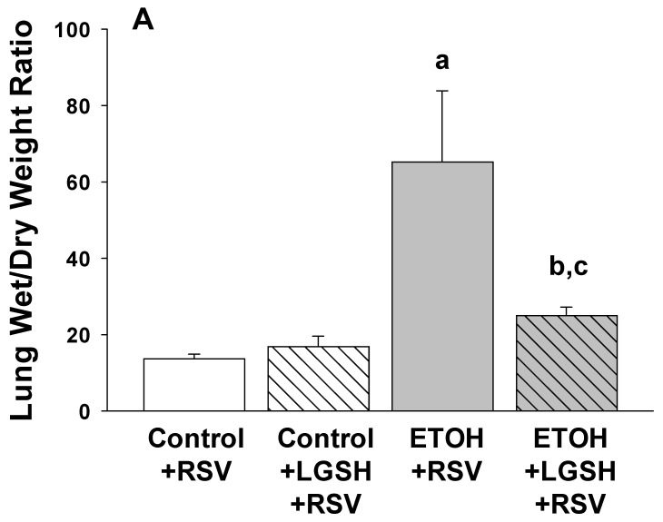
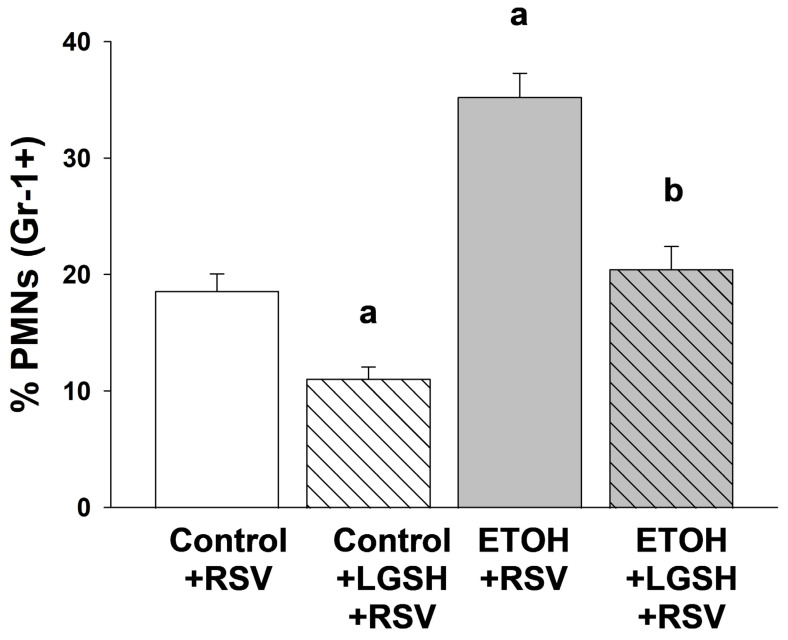
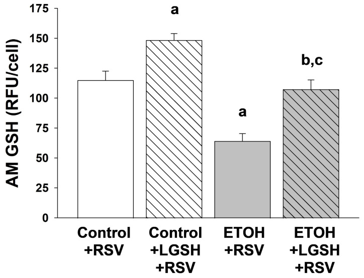
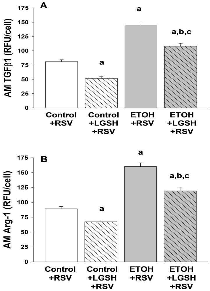
We previously reported that maternal alcohol use increased the risk of sepsis in premature and term newborns. In the neonatal mouse, fetal ethanol (ETOH) exposure depleted the antioxidant glutathione (GSH), which promoted alveolar macrophage (AM) immunosuppression and respiratory syncytial virus (RSV) infections. In this study, we explored if oral liposomal GSH (LGSH) would attenuate oxidant stress and RSV infections in the ETOH-exposed mouse pups. C57BL/6 female mice were pair-fed a liquid diet with 25% of calories from ethanol or maltose-dextrin. Postnatal day 10 pups were randomized to intranasal saline, LGSH, and RSV. After 48 h, we assessed oxidant stress, AM immunosuppression, pulmonary RSV burden, and acute lung injury. Fetal ETOH exposure increased oxidant stress threefold, lung RSV burden twofold and acute lung injury threefold. AMs were immunosuppressed with decreased RSV clearance. However, LGSH treatments of the ETOH group normalized oxidant stress, AM immune phenotype, the RSV burden, and acute lung injury. These studies suggest that the oxidant stress caused by fetal ETOH exposure impaired AM clearance of infectious agents, thereby increasing the viral infection and acute lung injury. LGSH treatments reversed the oxidative stress and restored AM immune functions, which decreased the RSV infection and subsequent acute lung injury.

Keywords: alveolar macrophage; fetal ethanol exposure; liposomal glutathione; oxidant stress; respiratory syncytial virus.


Conflict of interest statement

The authors declare no conflict of interest.

Figures

Figure 1
Figure 1
In utero ethanol (ETOH) exposure impaired in vivo alveolar macrophage (AM) phagocytosis of respiratory syncytial virus (RSV) but restored by enteral liposomal glutathione (LGSH) treatments. After breeding, the female mice were randomized to an experimental isocaloric liquid diet that contained either 25% ethanol (ETOH)-derived calories or 25% maltose–dextrin-derived calories. The diet was continued throughout pregnancy and after spontaneous term delivery. Pups were kept with their respective dams and allowed to nurse ad libitum. On postnatal day 10 (P10), pups from the control-fed dam and the ETOH-fed dam were then randomized to an oral gavage containing either liposomal glutathione (LGSH) (20 μL, 1.7 mg of L-glutathione) or saline (20 μL). All pups were then given intranasal injections of respiratory syncytial virus (RSV) (Nanoliter Injector; 20 µL; each nasal nare; 2 × 10⁵ PFU) before they were returned to their respective dams. After 24 h, pups received an additional dose of ±LGSH (or saline) by gavage. All pups were then euthanized for analyses after 48 h, and the alveolar macrophages (AMs) from the pups were isolated from the multiple BALs and pooled per experimental group and litter. For assessment of RSV phagocytosis by the AM in vivo, we evaluated whole-cell RSV via fluorescent immunostaining (1 h; a 1:100 dilution; Santa Cruz Biotechnology, Inc., Santa Cruz, CA, USA). Background fluorescence of unstained AMs was used to account for autofluorescence and subtracted from the RFUs obtained for the RSV phagocytosis. The RSV burden was calculated relative to the RSV fluorescence for AMs from the control + RSV group. N = 5 litters for each group. a p = 0.05 when compared to the control + RSV group; b p ≤ 0.05 when compared to the ETOH + RSV group.
Figure 2
Figure 2
In utero ethanol (ETOH) exposure increased the respiratory syncytial virus (RSV) burden in the bronchoalveolar lavage (BAL, A) and lung tissue (B), but both were normalized by oral liposomal glutathione (LGSH) treatments. All pups were euthanized for analyses after 48 h of RSV delivery and the lungs were then serially lavaged via the catheter with 40 µL sterile saline (5 times) to remove the fluid and cells from the airspace. To determine the RSV burden in the airways and alveolar space, the multiple BALs from pups were pooled per experimental group and litter. The pooled BAL was serially diluted in phosphate-buffered saline, plated for determination of RSV growth. The RSV burden in the BAL (A) is presented as percentage of control plaque forming units/mL (PFU/mL). To determine the RSV burden in the lung, the frozen right upper lung lobe was weighed, sterile phosphate-buffered saline (10 times the tissue weight) was added, and then the lobe homogenized on ice. After the samples were centrifuged (2000× g; 10 min; 4 °C), the supernatants were serially diluted in phosphate-buffered saline and plated for determination of RSV growth. For the whole lung (B), RSV is presented as percentage of control of PFU/g lung tissue. N = 5 litters for each group. a p = 0.05 when compared to the control + RSV group; b p ≤ 0.05 when compared to the ETOH + RSV group.
Figure 3
Figure 3
The amplified Respiratory syncytial virus (RSV) infection in the pup after in utero ethanol (ETOH) exposure resulted in increased acute lung injury, as defined by lung wet/dry weight ratio (A) and whole-lung myeloperoxidase (MPO) concentration (B), but acute lung injury was normalized by oral liposomal glutathione (LGSH) treatments. The flash-frozen right upper lobe lung samples from the neonatal pups were weighed (designated wet weight) and then reweighed after desiccation by overnight incubation at 70 °C (designated dry weight). The lung wet/dry weight ratio was determined as a marker of acute lung injury (A). The flash-frozen lung tissue was also evaluated for the inflammatory PMN marker myeloperoxidase (MPO) with a commercially available ELISA (product # MBS2702122, MyBioSource, Inc., San Diego, CA, USA). MPO concentrations (ng/mL) were normalized to the corresponding lung sample wet/dry weight (B). N = 5 litters for each group. a p = 0.05 when compared to the control + respiratory syncytial virus (RSV) group; b p ≤ 0.05 when compared to the ETOH + RSV group; c denotes p ≤ 0.05 when compared to the control + LGSH + RSV group.
Figure 3
Figure 3
The amplified Respiratory syncytial virus (RSV) infection in the pup after in utero ethanol (ETOH) exposure resulted in increased acute lung injury, as defined by lung wet/dry weight ratio (A) and whole-lung myeloperoxidase (MPO) concentration (B), but acute lung injury was normalized by oral liposomal glutathione (LGSH) treatments. The flash-frozen right upper lobe lung samples from the neonatal pups were weighed (designated wet weight) and then reweighed after desiccation by overnight incubation at 70 °C (designated dry weight). The lung wet/dry weight ratio was determined as a marker of acute lung injury (A). The flash-frozen lung tissue was also evaluated for the inflammatory PMN marker myeloperoxidase (MPO) with a commercially available ELISA (product # MBS2702122, MyBioSource, Inc., San Diego, CA, USA). MPO concentrations (ng/mL) were normalized to the corresponding lung sample wet/dry weight (B). N = 5 litters for each group. a p = 0.05 when compared to the control + respiratory syncytial virus (RSV) group; b p ≤ 0.05 when compared to the ETOH + RSV group; c denotes p ≤ 0.05 when compared to the control + LGSH + RSV group.
Figure 4
Figure 4
Respiratory syncytial virus (RSV) superimposed on in utero ethanol (ETOH) exposure increased polymorphonuclear leukocyte (PMN) migration into the alveolar space, but was attenuated by oral liposomal glutathione (LGSH) treatments. Immunostaining for the cell surface marker (Gr-1) was used to differentiate AM from the polymorphonuclear leukocytes (PMNs) that had migrated into the alveolar space. Cells retrieved from the lavage were plated, fixed, and incubated with the GR-1 primary antibody in a 1:100 dilution (Santa Cruz Biotechnology, Inc., Santa Cruz, CA, USA) for 1 h. After the slides were washed three times with phosphate-buffered saline over 5 min, the secondary antibody (anti-goat IgG) was added in a 1:200 dilution and further incubated for 45 min. Cellular fluorescence was quantified using fluorescence microscopy via ImagePro Plus for Windows Version 4.5 and presented as mean relative fluorescence units per cell (RFUS/cell) ± S.E.M. tallied from at least 25 cells/litter. To correct for autofluorescence, the background fluorescence of unstained AMs was subtracted from the RFUs obtained for each analysis. N = 5 litters for each group. a p = 0.05 when compared to the control + respiratory syncytial virus (RSV) group; b p ≤ 0.05 when compared to the ETOH + RSV group.
Figure 5
Figure 5
In utero ethanol (ETOH) exposure increased oxidant stress, as defined by a decrease in the plasma antioxidant capacity (AOC; A) and an increase in DNA oxidation (8OH-dG; B), but this oxidant stress was attenuated by oral liposomal glutathione (LGSH) treatments. After euthanasia with intraperitoneal sodium pentobarbital, blood samples were obtained from all pups via cardiac puncture and the samples pooled per experimental group and litter. Samples were spun and stored at −80 °C until batch analyses. Total plasma AOC (A) was measured via colorimetric assay (MAK187, Sigma-Aldrich, St. Louis, MO, USA) and 8-OHdG (B) was measured by ELISA (DNA Damage Competitive ELISA Kit, Life Technologies Corporation, Carlsbad, CA, USA). N = 5 litters for each group. a p = 0.05 when compared to the control + RSV group; b p ≤ 0.05 when compared to the ETOH + respiratory syncytial virus (RSV) group; c p ≤ 0.05 when compared to the control + LGSH + RSV group.
Figure 6
Figure 6
In utero ethanol (ETOH) exposure increased alveolar macrophage (AM) oxidant stress, as defined by a decrease in the glutathione (GSH) pool, but the GSH pool in the AM was restored by oral liposomal glutathione (LGSH) treatments. The freshly isolated AMs were plated and fixed with 3.7% paraformaldehyde before permeabilization with ice-cold methanol. The antioxidant GSH pool in the AMs was evaluated by whole-cell GSH via fluorescent immunostaining (1:100 dilution; Abcam, Inc®, Boston, MA, USA). Cellular fluorescence was quantified using fluorescence microscopy via ImagePro Plus for Windows version 4.5 [37] and is presented as mean relative fluorescence units per cell (RFUS/cell) ± S.E.M. as tallied from at least 25 cells/litter. To correct for autofluorescence, the background fluorescence of unstained AMs was subtracted from the RFUs obtained for each analysis. N = 5 litters for each group. a p = 0.05 when compared to the control + respiratory syncytial virus (RSV) group; b p ≤ 0.05 when compared to the ETOH + RSV group; c p ≤ 0.05 when compared to the control + LGSH + RSV group.
Figure 7
Figure 7
In utero ethanol (ETOH) exposure resulted in alveolar macrophage (AM) immunosuppression, as defined by an increase in transforming growth factor β1 (TGFβ1) (A) and arginase 1 (Arg-1) (B) expression, but oral liposomal glutathione (LGSH) treatments attenuated the AM immunosuppression. AM expression of TGFβ1 (A) and Arg-1 (B) were used as markers of immunosuppression. Cells were incubated with the primary antibody in a 1:100 dilution (Santa Cruz Biotechnology, Inc., Santa Cruz, CA, USA) for 1 h. After the slides were washed three times with phosphate-buffered saline over 5 min, the secondary antibody (anti-goat IgG) was added in a 1:200 dilution and further incubated for 45 min. Cellular fluorescence was quantified using fluorescence microscopy via ImagePro Plus for Windows version 4.5 [38] and is presented as mean relative fluorescence units per cell (RFUS/cell) ± S.E.M. tallied from at least 25 cells/litter. To correct for autofluorescence, the background fluorescence of unstained AMs was subtracted from the RFUs obtained for each analysis. N = 5 litters for each group. a p = 0.05 when compared to the control + respiratory syncytial virus (RSV) group; b p ≤ 0.05 when compared to the ETOH + RSV group; c p ≤ 0.05 when compared to the control + LGSH + RSV group.

Similar articles

Cited by

References

    1. Gauthier T.W. Prenatal Alcohol Exposure and the Developing Immune System. Alcohol Res. Curr. Rev. 2015;37:279–285. - PMC - PubMed
    1. Gauthier T.W., Guidot D.M., Kelleman M.S., McCracken C.E., Brown L.A. Maternal Alcohol Use During Pregnancy and Associated Morbidities in Very Low Birth Weight Newborns. Am. J. Med. Sci. 2016;352:368–375. doi: 10.1016/j.amjms.2016.06.019. - DOI - PMC - PubMed
    1. Henriksen T.B., Hjollund N.H., Jensen T.K., Bonde J.P., Andersson A.M., Kolstad H., Ernst E., Giwercman A., Skakkebaek N.E., Olsen J. Alcohol consumption at the time of conception and spontaneous abortion. Am. J. Epidemiol. 2004;160:661–667. doi: 10.1093/aje/kwh259. - DOI - PubMed
    1. Kesmodel U., Wisborg K., Olsen S.F., Henriksen T.B., Secher N.J. Moderate alcohol intake during pregnancy and the risk of stillbirth and death in the first year of life. Am. J. Epidemiol. 2002;155:305–312. doi: 10.1093/aje/155.4.305. - DOI - PubMed
    1. Kesmodel U., Wisborg K., Olsen S.F., Henriksen T.B., Secher N.J. Moderate alcohol intake in pregnancy and the risk of spontaneous abortion. Alcohol Alcohol. 2002;37:87–92. doi: 10.1093/alcalc/37.1.87. - DOI - PubMed

LinkOut - more resources