. 2024 Jan 29;12(2):309.
doi: 10.3390/biomedicines12020309.

Treatment with Cobra Venom Factor Decreases Ischemic Tissue Damage in Mice

Affiliations

Treatment with Cobra Venom Factor Decreases Ischemic Tissue Damage in Mice

Sharon O Azubuike-Osu et al. Biomedicines. .

Abstract

Tissue ischemia, caused by the blockage of blood vessels, can result in substantial damage and impaired tissue performance. Information regarding the functional contribution of the complement system in the context of ischemia and angiogenesis is lacking. To investigate the influence of complement activation and depletion upon femoral artery ligation (FAL), Cobra venom factor (CVF) (that functionally resembles C3b, the activated form of complement component C3) was applied in mice in comparison to control mice. Seven days after induction of muscle ischemia through FAL, gastrocnemius muscles of mice were excised and subjected to (immuno-)histological analyses. H&E and apoptotic cell staining (TUNEL) staining revealed a significant reduction in ischemic tissue damage in CVF-treated mice compared to controls. The control mice, however, exhibited a significantly higher capillary-to-muscle fiber ratio and a higher number of proliferating endothelial cells (CD31+/CD45-/BrdU+). The total number of leukocytes (CD45+) substantially decreased in CVF-treated mice versus control mice. Moreover, the CVF-treated group displayed a shift towards the M2-like anti-inflammatory and regenerative macrophage phenotype (CD68+/MRC1+). In conclusion, our findings suggest that treatment with CVF leads to reduced ischemic tissue damage along with decreased leukocyte recruitment but increased numbers of M2-like polarized macrophages, thereby enhancing tissue regeneration, repair, and healing.

Keywords: angiogenesis; cobra venom factor; complement system; inflammation; ischemia; leukocytes; macrophage polarization; macrophages; vascular occlusion.


Conflict of interest statement

The authors declare no conflicts of interest.

Figures
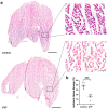
Figure 1
Figure 1
Tissue damage is reduced in CVF-treated mice. (a) Representative H&E-stained images of gastrocnemius muscles (left) with magnifications (right) of the areas shown in the black boxes of control mice (upper images) and cobra venom factor-treated mice (lower images) taken 7 days aFAL. (b) Scatter plot presenting the area of ischemic tissue damage (%) in control and CVF-treated mice 7 days after femoral artery ligation (aFAL). One complete sectional area was assessed per mouse per group. The data represent means ± SEM, with n = 5 per group. *** p ≤ 0.001 (control vs. CVF) determined by unpaired Student’s t-tests. The scale bars represent 1000 µm (overview) and 100 µm (detail).
Figure 2
Figure 2
TUNEL staining revealed decreased apoptosis in CVF-treated mice compared to control mice. (a) The scatter plot depicts the apoptotic area (%) in control and CVF-treated mice 7 days after femoral artery ligation (aFAL). Analysis was conducted on one complete sectional area per mouse per group. The data presented are means ± SEM, with n = 5 per group. ** p ≤ 0.01 (control vs. CVF) determined by unpaired Student’s t-tests. (b) Magnified images (right) of the areas shown in the white boxes of the representative images (left) of TUNEL-stained gastrocnemius muscles from control mice (upper images) and CVF-treated mice (lower images) showing apoptotic cells (green) at 7 days aFAL. Scale bars: 1000 µm (overview), 100 µm (detail).
Figure 3
Figure 3
CVF treatment led to reduced capillarity. Scatter plots illustrate the number of (a) endothelial cells (CD31+/CD45) and (b) proliferating endothelial cells (CD31+/CD45/BrdU+) per muscle fiber of ischemic gastrocnemius muscles in control and CVF-treated mice 7 days after femoral artery ligation (aFAL). The data presented are means ± SEM, with n = 5 per group. A defined ischemic area (1.5 mm2) of muscle tissue was analyzed per mouse. **** p ≤ 0.0001 (control vs. CVF) determined by unpaired Student’s t-tests. (c) Representative immunofluorescence images of analyzed ischemic gastrocnemius muscles from control (upper images) and CVF-treated mice (lower images) at 7 days aFAL. Single channel pictures (small images on the right) and merged pictures (large images on the left) show endothelial cells (CD31 in gray), proliferating cells (BrdU in red), leukocytes (CD45 in green), and the nuclei (DAPI in blue). Scale bars: 50 µm.
Figure 4
Figure 4
CVF treatment reduced leukocyte infiltration in ischemic gastrocnemius muscles. (a) The scatter plot illustrates the number of leukocytes (CD45+) per square millimeter (mm2) in ischemic gastrocnemius muscles isolated 7 days after femoral artery ligation (aFAL). The data presented are means ± SEM, with n = 5 per group. Analysis was conducted on a defined ischemic area (1.5 mm2) of muscle tissue per mouse. *** p ≤ 0.001 (control vs. CVF) determined by unpaired Student’s t-tests. (b) Representative immunofluorescence images of analyzed gastrocnemius muscles from control (left image) and CVF-treated mice (right image) at 7 days aFAL. Leukocytes in single-channel pictures (lower images) and merged pictures (upper images) were labeled with CD45 antibodies (green) and the nuclei were labeled with DAPI (blue). Scale bars: 100 µm.
Figure 5
Figure 5
CVF treatment promotes M2-like polarization in ischemic muscle tissue. The scatter plot displays (a) the total number of macrophages (CD68+) per square millimeter (mm2) and the percentage of (b) M1-like polarized macrophages (CD68+/MRC1) and (c) M2-like polarized macrophages (CD68+/MRC1+) in ischemic gastrocnemius muscle tissue at 7 days after femoral artery ligation (aFAL). Data are shown as means ± SEM, with n = 5 per group. ** p ≤ 0.01 and **** p ≤ 0.0001 (control vs. CVF) determined by unpaired Student’s t-tests. (d) Representative immunofluorescence images of analyzed ischemic gastrocnemius muscles from control (upper image) and CVF-treated mice (lower image) at 7 days aFAL. In single-channel pictures (small images on the right) and merged pictures (large images on the left), macrophages were labeled with CD68 antibody (green) and M2-like polarized macrophages were labeled with MRC1 antibody (red). Nuclei were labeled in merged images with DAPI (blue). Scale bars: 50 µm.
Figure 6
Figure 6
The connection between arteriogenesis and angiogenesis. The left picture shows a healthy leg with pre-existing collateral arteries. The leg in the middle displays an occluded main artery with ischemic tissue damage and a capillary network in the lower leg as a result of the occlusion in the upper leg. The leg on the right side shows the effect of arteriogenesis to decrease the extent of ischemia in the lower leg (Image from Chillo et al. [33]).

Similar articles

References

    1. Sao C.W., Aday A.W., Almarzooq Z.I., Anderson C.A., Arora P., Avery C.L., Baker-Smith C.M., Beaton A.Z., Boehme A.K., Buxton A.E., et al. Heart Disease and Stroke Statistics—2023 Update: A Report From the American Heart Association. Circulation. 2023;147:e93–e621. - PubMed
    1. Hausenloy D.J., Yellon D.M. Myocardial ischemia-reperfusion injury: A neglected therapeutic target. J. Clin. Investig. 2013;123:92–100. doi: 10.1172/JCI62874. - DOI - PMC - PubMed
    1. Ricklin D., Hajishengallis G., Yang K., Lambris J.D. Complement: A key system for immune surveillance and homeostasis. Nat. Immunol. 2010;11:785–797. doi: 10.1038/ni.1923. - DOI - PMC - PubMed
    1. Merle N.S., Noe R., Halbwachs-Mecarelli L., Fremeaux-Bacchi V., Roumenina L.T. Complement System Part II: Role in Immunity. Front. Immunol. 2015;6:257. doi: 10.3389/fimmu.2015.00257. - DOI - PMC - PubMed
    1. Hajishengallis G., Reis E.S., Mastellos D.C., Ricklin D., Lambris J.D. Novel mechanisms and functions of complement. Nat. Immunol. 2017;18:1288–1298. doi: 10.1038/ni.3858. - DOI - PMC - PubMed

Grants and funding