. 2024 Feb 15;16(4):792.
doi: 10.3390/cancers16040792.

The Immunomodulatory Effects of Fluorescein-Mediated Sonodynamic Treatment Lead to Systemic and Intratumoral Depletion of Myeloid-Derived Suppressor Cells in a Preclinical Malignant Glioma Model

Affiliations

The Immunomodulatory Effects of Fluorescein-Mediated Sonodynamic Treatment Lead to Systemic and Intratumoral Depletion of Myeloid-Derived Suppressor Cells in a Preclinical Malignant Glioma Model

Serena Pellegatta et al. Cancers (Basel). .

Abstract

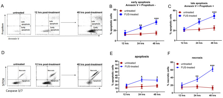
Fluorescein-mediated sonodynamic therapy (FL-SDT) is an extremely promising approach for glioma treatment, resulting from the combination of low-intensity focused ultrasound (FUS) with a sonosensitizer. In the present study, we evaluated the efficacy and immunomodulation of SDT with fluorescein as the sonosensitizer in immunocompetent GL261 glioma mice for the first time. In vitro studies demonstrated that the exposure of GL261 cells to FL-SDT induced immunogenic cell death and relevant upregulation of MHC class I, CD80 and CD86 expression. In vivo studies were then performed to treat GL261 glioma-bearing mice with FL-SDT, fluorescein alone, or FUS alone. Perturbation of the glioma-associated macrophage subset within the immune microenvironment was induced by all the treatments. Notably, a relevant depletion of myeloid-derived suppressor cells (MDSCs) and concomitant robust infiltration of CD8+ T cells were observed in the SDT-FL-treated mice, resulting in a significant radiological delay in glioma progression and a consequent improvement in survival. Tumor control and improved survival were also observed in mice treated with FL alone (median survival 41.5 days, p > 0.0001 compared to untreated mice), reflecting considerable modulation of the immune microenvironment. Interestingly, a high circulating lymphocyte-to-monocyte ratio and a very low proportion of MDSCs were predictive of better survival in FL- and FL-SDT-treated mice than in untreated and FUS-treated mice, in which elevated monocyte and MDSC frequencies correlated with worse survival. The immunostimulatory potential of FL-SDT treatment and the profound modulation of most immunosuppressive components within the microenvironment encouraged the exploration of the combination of FL-SDT with immunotherapeutic strategies.

Keywords: CD8 infiltrating T cells; MDSC; fluorescein; glioma; immune microenvironment; mouse model; sonodynamic therapy; ultrasound.


Conflict of interest statement

The authors declare no conflicts of interest.

Figures

Figure 1
Figure 1
(A) Representative flow cytometry dot plots of Annexin V-PI-stained GL261-NS cells. GL261-NS cells incubated with FL only or treated with FL-SDT were analyzed for cell death. Each plot shows cells positive for Annexin V only (LR), positive for PI only (region UL), and negative for both (region LL), representing early apoptotic, late apoptotic, and live cells, respectively. (B,C) The graphs show the percentage of early (B) and late (C) apoptotic cells measured after 12, 24, and 48 h (red line, FL-only or “untreated”; blue line, FL-SDT-treated or “FUS-treated”). (D) Representative flow cytometry dot plots for Caspase 3/Caspase 7 indicating live cells (LL), late apoptotic cells (LR), and necrotic cells (UR). (E,F) The graphs show the percentages of apoptotic (E) and necrotic (F) cells measured after 12, 24, and 48 h (red line, FL-only; blue line, FL-SDT-treated cells). (* p < 0.01, ** p < 0.005, *** p < 0.0001; FL + SDT vs. FL only). The data from three experiments are presented as the mean ± SD.
Figure 2
Figure 2
(A,C,E) Representative flow cytometry dot plots showing the expression of MHCI (A), costimulatory CD80 (C), and CD86 (E) on GL261-NS cells treated with FL-SDT compared with that on cells incubated with FL. (B,D,F) Bar graphs showing the percentage of positive cells as assessed by flow cytometry at 12, 24, and 48 h after FUS treatment compared with that of untreated but FL-preincubated GL261-NS cells. (GL) Bar graphs showing the relative expression of different NKG2D ligands at different time points after FL-SDT treatment compared to that in untreated cells (* p < 0.01, ** p < 0.005, *** p < 0.0001; FL + SDT vs. FL only). The data from three experiments are presented as the mean ± SD.
Figure 3
Figure 3
(AD) Representative MRI images performed at three different time points with T2-weighted images (T2-wi) and T1-weighted images (T1-wi) with contrast medium injection showing the pattern of tumor progression in untreated control mice (A), treated with FUS only (B), sodium fluorescein (NaFL) only (C), and FL-SDT (D). (E) Time courses showing the tumor diameters of the mice in (AD) measured via MRI. (F) K–M survival curves of untreated mice and mice treated with FUS only, NaFL only, or NaFL-SDT (n = 6/group). Overall survival statistical analysis was performed using the Mantel–Cox log-rank test.
Figure 4
Figure 4
(A) Representative dot plots showing the immune cell infiltration assessed using CD11b and CD45 as markers. (B) Bar graphs showing the percentages of the different subsets of infiltrating immune cells, including resting and activated tumor-associated macrophages and lymphocytes. (C) Percentage of infiltrating MDSCs assessed by flow cytometry as CD11b+Gr1+ on the CD45+ gate in untreated and treated glioma tissues at 14, 21, and 28 days after tumor implantation. (D) Percentage of infiltrating CD8+ T cells assessed by flow cytometry as CD3+ CD8+ cells in the CD45high gate in untreated and treated glioma tissues at 14, 21, and 28 days after tumor implantation. (E) Representative dot plot showing the lymphocyte and monocyte counts on SSCs and the percentage of peripheral MDSCs (CD11b+ Gr+) in untreated and treated mice. (F) Time course of peripheral MDSCs in all the groups at the three time points evaluated for intratumoral quantification and later time points for FL−only and FL−SDT. (F,G) Time course of peripheral T cells in all the groups at the three time points evaluated for intratumoral quantification and later time points for FL−only and FL−SDT. (* p < 0.01, ** p < 0.005, *** p < 0.0001). The data from three experiments are presented as the mean ± SD.

References

    1. Wang X.-X., Cao H., Zhai Y., Deng S.-Z., Chao M., Hu Y., Mou Y., Guo S., Zhao W., Li C., et al. Immune gene signatures and immunotypes in immune microenvironment are associated with glioma prognose. Front. Immunol. 2022;13:823910. doi: 10.3389/fimmu.2022.823910. - DOI - PMC - PubMed
    1. Najem H., Khasraw M., Heimberger A.B. Immune microenvironment landscape in CNS tumors and role in responses to immunotherapy. Cells. 2021;10:2032. doi: 10.3390/cells10082032. - DOI - PMC - PubMed
    1. Musca B., Russo M.G., Tushe A., Magri S., Battaggia G., Pinton L., Bonaudo C., Della Puppa A., Mandruzzato S. The immune cell landscape of glioblastoma patients highlights a myeloid-enriched and immune suppressed microenvironment compared to metastatic brain tumors. Front. Immunol. 2023;14:1236824. doi: 10.3389/fimmu.2023.1236824. - DOI - PMC - PubMed
    1. Anderson R.C.E., Anderson D.E., Elder J.B., Brown M.D., Mandigo C.E., Parsa A.T., Goodman R.R., McKhann G.M., Sisti M.B., Bruce J.N. Lack of B7 expression, not human leukocyte antigen expression, facilitates immune evasion by human malignant gliomas. Neurosurgery. 2007;60:1129–1136; discussion 1136. doi: 10.1227/01.NEU.0000255460.91892.44. - DOI - PubMed
    1. Roesch S., Rapp C., Dettling S., Herold-Mende C. When immune cells turn bad—tumor-associated microglia/macrophages in glioma. Int. J. Mol. Sci. 2018;19:436. doi: 10.3390/ijms19020436. - DOI - PMC - PubMed