Review
. 2024 Feb 15;13(4):1101.
doi: 10.3390/jcm13041101.

Sacroplasty for Sacral Insufficiency Fractures: Narrative Literature Review on Patient Selection, Technical Approaches, and Outcomes

Affiliations
Review

Sacroplasty for Sacral Insufficiency Fractures: Narrative Literature Review on Patient Selection, Technical Approaches, and Outcomes

Manjot Singh et al. J Clin Med. .

Abstract

Sacral insufficiency fractures commonly affect elderly women with osteoporosis and can cause debilitating lower back pain. First line management is often with conservative measures such as early mobilization, multimodal pain management, and osteoporosis management. If non-operative management fails, sacroplasty is a minimally invasive intervention that may be pursued. Candidates for sacroplasty are patients with persistent pain, inability to tolerate immobilization, or patients with low bone mineral density. Before undergoing sacroplasty, patients' bone health should be optimized with pharmacotherapy. Anabolic agents prior to or in conjunction with sacroplasty have been shown to improve patient outcomes. Sacroplasty can be safely performed through a number of techniques: short-axis, long-axis, coaxial, transiliac, interpedicular, and balloon-assisted. The procedure has been demonstrated to rapidly and durably reduce pain and improve mobility, with little risk of complications. This article aims to provide a narrative literature review of sacroplasty including, patient selection and optimization, the various technical approaches, and short and long-term outcomes.

Keywords: balloon-assisted; coaxial; interpedicular; long-axis; sacral insufficiency fractures; sacroplasty techniques; short-axis; transiliac.


Conflict of interest statement

B.G.D. discloses consulting fees from Spineart, Clariance, and Spinevision. J.K.C. discloses consulting fees from Stryker, Globus, Lifespan, CTL Amedica, Spineart, and Neuromonitoring Associates. A.H.D. discloses the following, receives royalties from Spineart and Stryker, consulting fees from Medtronic, research support from Alphatec, Medtronic, and Orthofix, and fellowship support from Medtronic. The other authors have nothing to disclose.

Figures

Figure 1
Figure 1
“ Classification of fragility fractures of the pelvis. (a) FFP Type Ia: unilateral anterior pelvic ring disruption. (b) FFP Type Ib: bilateral anterior pelvic ring disruption. (c) FFP Type IIa: dorsal non-displaced posterior injury only. (d) FFP Type IIb: sacral crush with anterior disruption. (e) FFP Type IIc: non-displaced sacral, sacroiliac or iliac fracture with anterior disruption. (f) FFP Type IIIa: displaced unilateral ilium fracture and anterior disruption. (g) FFP Type IIIb: displaced unilateral sacroiliac disruption and anterior disruption. (h) FFP Type IIIc: displaced unilateral sacral fracture together with anterior disruption. (i) FFP Type IVa: bilateral iliac fractures or bilateral sacroiliac disruptions together with anterior disruption. (j) FFP Type IVb: spinopelvic dissociation with anterior disruption. (k) FFP Type IVc: combination of different posterior instabilities together with anterior disruption” by Rommens et al. (Accessed 12 December 2023 at https://doi.org/10.1007/s00776-014-0653-9). Licensed under CC BY-NC-ND 4.0 © 2014 The Japanese Orthopaedic Association. To view a copy of this license, visit https://creativecommons.org/licenses/by-nc-nd/4.0/ (Red color indicates fracture lines).
Figure 2
Figure 2
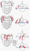
Bakker classification for sacral insufficiency fractures adapted from “Type A-fractures of the sacral ala: A1 with bone bruise (MRI) without a visible fracture line in the CT-scan; A2 deformation of the anterior cortical bone without a cortical disruption; and A3 anterolateral rim fracture of the ala with up to 1 cm distance in the direction of the medial sacroiliac joint.” (top), “ Type B fractures of the sacral ala: B1 fracture parallel to the sacroiliac joint; B2 fracture involving the sacroiliac joint; and B3 fracture with an involvement of the neural foramina or the spinal canal.” (middle), and “ Type C- or corpus-fractures: C1 fracture moves from anterior cortex dorsally or into the sacroiliac joint; C2 fracture with an unilateral involvement of the neural foramina or the spinal canal; and C3 is unstable and represents bilaterally sagittal fractures combined with a transverse lesion.” (bottom) by Bakker et al. (https://doi.org/10.3340/jkns.2017.0188). Original figures licensed under CC BY-NC-ND 4.0© 2018 The Korean Neurosurgical Society. To view a copy of this license, visit https://creativecommons.org/licenses/by-nc-nd/4.0/ (Red indicates fracture lines).
Figure 3
Figure 3
Sacroplasty surgical techniques: (A) bilateral short axis, (B) bilateral long axis, (C) coaxial vision, (D) transiliac, (E) interpedicular, (F) balloon-assisted. (Blue color denotes the site of cement deposition. Red color denotes the space created by balloon assistance).
Figure 4
Figure 4
Pre-operative, intraoperative, and post-operative imaging for patient case 2. Pre-operative anteroposterior (A) and lateral (B) lumbosacral spine radiographs demonstrated spondylosis of L3 over L4. Axial (C) and coronal (D) CT confirmed L3–4 dynamic spondylolisthesis and bilateral sacral alar insufficiency fractures. Axial I and sagittal (E,F) MRI STIR sequence of a related patient case showing edema at the fracture site. Intraoperative imaging during bilateral long-view sacroplasty demonstrated (G) successful ball tip probe cannulation of the right sacral ala without anterior penetration of the sacrum on anteroposterior inlet view, (H) Jamshidi cannulation down the right SI joint with no joint penetration on 25° right oblique view, (I) Jamshidi directed towards the anterior vertebral body with appropriate trajectory on lateral view, (J) cement filling the left sacral ala without joint or foraminal penetration on left oblique view, and (K) no final anterior or superior extravasation of cement on lateral view. Post-operative anteroposterior (L) and lateral (M) lumbosacral spine radiographs re-demonstrated spondylosis of L3 over L4 and good distribution of the cement without extravasation.
Figure 5
Figure 5
Pre-operative, intraoperative, and post-operative imaging for patient case 1. Pre-operative anteroposterior (A) and lateral (B) lumbosacral spine radiographs demonstrated degenerative spondylolitic changes of the lumbar vertebrae. Axial (C) and coronal (D) CT confirmed bilateral sacral fractures centered around S3. Intraoperative (EH) imaging during bilateral short-view sacroplasty revealed adequate cement deposition at the fracture site. Post-operative anteroposterior (I) and lateral (J) lumbosacral spine radiographs showed good distribution of the cement without extravasation.

Similar articles

Cited by

References

    1. Urits I., Orhurhu V., Callan J., Maganty N.V., Pousti S., Simopoulos T., Yazdi C., Kaye R.J., Eng L.K., Kaye A.D., et al. Sacral Insufficiency Fractures: A Review of Risk Factors, Clinical Presentation, and Management. Curr. Pain Headache Rep. 2020;24:10. doi: 10.1007/s11916-020-0848-z. - DOI - PubMed
    1. Blake S.P., Connors A.M. Sacral insufficiency fracture. Br. J. Radiol. 2004;77:891–896. doi: 10.1259/bjr/81974373. - DOI - PubMed
    1. Babayev M., Lachmann E., Nagler W. The controversy surrounding sacral insufficiency fractures: To ambulate or not to ambulate? Am. J. Phys. Med. Rehabil. 2000;79:404–409. doi: 10.1097/00002060-200007000-00014. - DOI - PubMed
    1. Tsiridis E., Upadhyay N., Giannoudis P.V. Sacral insufficiency fractures: Current concepts of management. Osteoporos. Int. 2006;17:1716–1725. doi: 10.1007/s00198-006-0175-1. - DOI - PubMed
    1. Pommersheim W., Huang-Hellinger F., Baker M., Morris P. Sacroplasty: A Treatment for Sacral Insufficiency Fractures. AJNR Am. J. Neuroradiol. 2003;24:1003–1007. - PMC - PubMed

LinkOut - more resources