. 2024 Feb 9;16(2):256.
doi: 10.3390/pharmaceutics16020256.

Repurposed Drugs That Activate Autophagy in Filarial Worms Act as Effective Macrofilaricides

Affiliations

Repurposed Drugs That Activate Autophagy in Filarial Worms Act as Effective Macrofilaricides

Denis Voronin et al. Pharmaceutics. .

Abstract

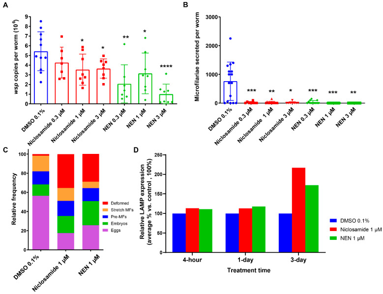
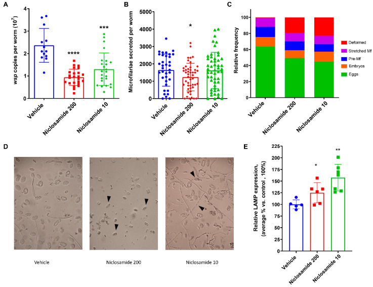
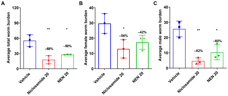
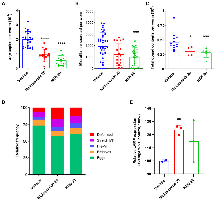
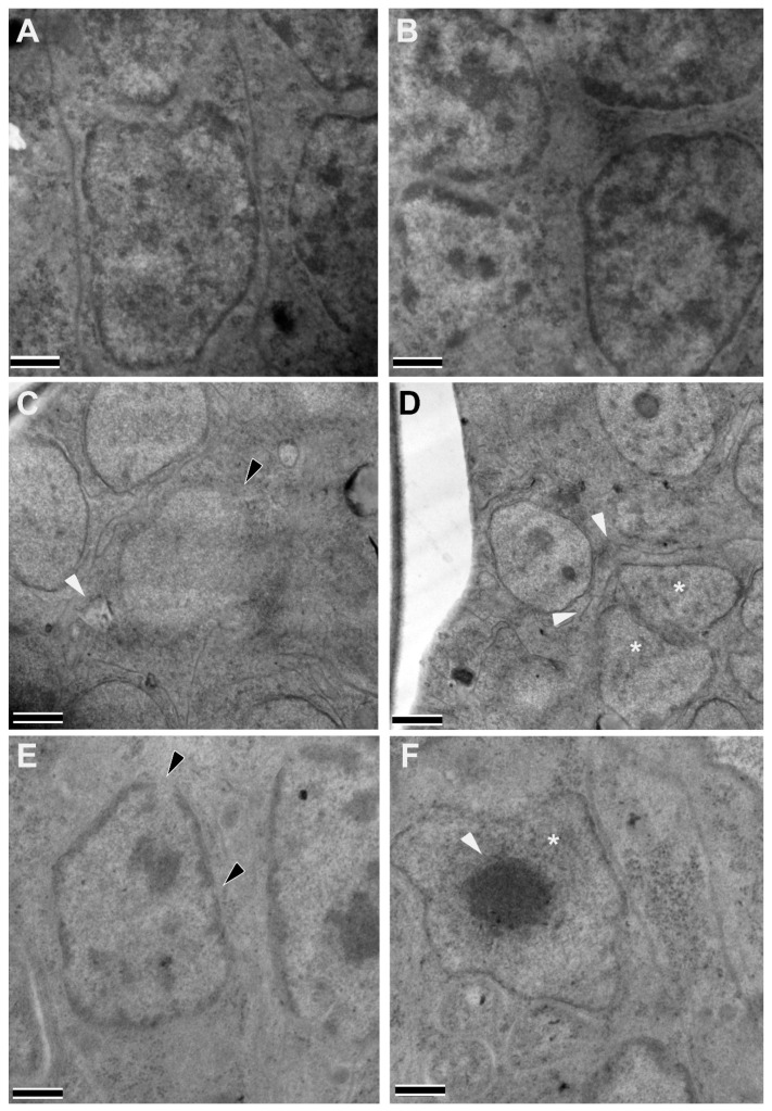
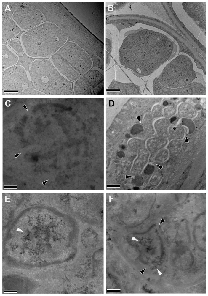
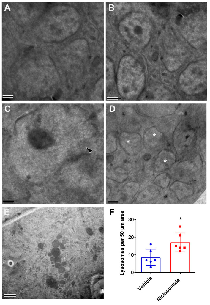
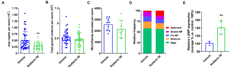
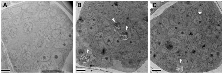
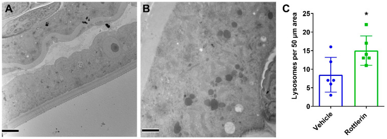
Onchocerciasis and lymphatic filariasis are two neglected tropical diseases caused by filarial nematodes that utilize insect vectors for transmission to their human hosts. Current control strategies are based on annual or biannual mass drug administration (MDA) of the drugs Ivermectin or Ivermectin plus Albendazole, respectively. These drug regimens kill the first-stage larvae of filarial worms (i.e., microfilariae) and interrupt the transmission of infections. MDA programs for these microfilaricidal drugs must be given over the lifetime of the filarial adult worms, which can reach 15 years in the case of Onchocerca volvulus. This is problematic because of suboptimal responses to ivermectin in various endemic regions and inefficient reduction of transmission even after decades of MDA. There is an urgent need for the development of novel alternative treatments to support the 2030 elimination goals of onchocerciasis and lymphatic filariasis. One successful approach has been to target Wolbachia, obligatory endosymbiotic bacteria on which filarial worms are dependent for their survival and reproduction within the human host. A 4-6-week antibiotic therapy with doxycycline, for example, resulted in the loss of Wolbachia that subsequently led to extensive apoptosis of somatic cells, germline, embryos, and microfilariae, as well as inhibition of fourth-stage larval development. However, this long-course regimen has limited use in MDA programs. As an alternative approach to the use of bacteriostatic antibiotics, in this study, we focused on autophagy-inducing compounds, which we hypothesized could disturb various pathways involved in the interdependency between Wolbachia and filarial worms. We demonstrated that several such compounds, including Niclosamide, an FDA-approved drug, Niclosamide ethanolamine (NEN), and Rottlerin, a natural product derived from Kamala trees, significantly reduced the levels of Wolbachia in vitro. Moreover, when these compounds were used in vivo to treat Brugia pahangi-infected gerbils, Niclosamide and NEN significantly decreased adult worm survival, reduced the release of microfilariae, and decreased embryonic development depending on the regimen and dose used. All three drugs given orally significantly reduced Wolbachia loads and induced an increase in levels of lysosome-associated membrane protein in worms from treated animals, suggesting that Niclosamide, NEN, and Rottlerin were effective in causing drug-induced autophagy in these filarial worms. These repurposed drugs provide a new avenue for the clearance of adult worms in filarial infections.

Keywords: Wolbachia; autophagy; filarial diseases; macrofilaricidal drugs; repurposed drugs.


Conflict of interest statement

The authors declare that they have no competing interests.

Figures

Figure 1
Figure 1
Treatment with Niclosamide and NEN in vitro reduced Wolbachia loads and worm fecundity and led to upregulation of lysosomal marker. (A) Wolbachia loads in treated and non-treated female worms were quantified using qPCR of the single copy wsp gene. (B) The number of microfilariae secreted from individual adult female worms was quantified over days 5–6 in culture. (C) Embryogram analyses using phase contrast microscopy were performed on individual female worms 6 days post-treatment, and the numbers of the various embryonic developmental stages (eggs, embryos, pre-microfilariae, stretched microfilariae, deformed embryos) in at least 200 events were determined. (D) Analysis of LAMP expression of treated worms (n = 2, per sample) by Western blot. LAMP values are normalized to tubulin expression. p-value < 0.05 (*), p-value < 0.01 (**) and p-value < 0.001 (***), p-value < 0.0001 (****).
Figure 2
Figure 2
In vivo treatment of infected gerbils with Niclosamide 200 mg/kg PO and 10 mg/kg IP reduced Wolbachia loads and induced deformity in worm progeny after a 10-day treatment regimen. (A) Wolbachia loads in female worms recovered from treated gerbils were quantified using qPCR of the single copy wsp gene. (B) The number of microfilariae secreted from individual adult female worms was quantified over days 5–6 in culture. (C) Embryogram analyses using phase contrast microscopy were performed on individual female worms 6 days post-treatment, and numbers for various embryonic developmental stages (eggs, embryos, pre-microfilariae, stretched microfilariae, deformed embryos) in at least 200 events were determined. (D) Representative phase contrast micrographs from the embryogram analyses. Black arrowheads demonstrate deformed progeny in treated worms. (E) Analysis of LAMP expression of treated worms by Western blot. LAMP expression is normalized to tubulin expression. p-value < 0.05 (*), p-value < 0.01 (**) and p-value < 0.001 (***), p-value < 0.0001 (****).
Figure 3
Figure 3
Treatment of infected gerbils with 20 mg/kg Niclosamide and NEN using the intraperitoneal route reduced worm burden after a 10-day treatment regimen. (A) Mean number of total worm burden per animal; (B) Mean number of female worm burden per animal; and (C) Mean number of male worm burden per animal. Necropsies were performed 4–6 days after final treatment, and worms were recovered from the peritoneum. p-value < 0.05 (*), p-value < 0.01 (**).
Figure 4
Figure 4
In vivo treatment of infected gerbils with Niclosamide 20 mg/kg and NEN 20 mg/kg IP affected Wolbachia loads, worm fecundity, and upregulated markers of autophagy after a 10-day treatment regimen. (A) Wolbachia loads in female worms recovered from treated gerbils were quantified using qPCR of the single copy wsp gene. (B) The number of microfilariae secreted from individual adult female worms was quantified over days 5–6 in culture. (C) Quantification of total gonad content in female worms. (D) Embryogram analyses using phase contrast microscopy were performed on individual female worms 6 days post-treatment, and the numbers of the various embryonic developmental stages (eggs, embryos, pre-microfilariae, stretched microfilariae, and deformed embryos) in at least 200 events were determined. (E) Analysis of LAMP expression of treated worms by Western blot. LAMP expression is normalized to tubulin expression. p-value < 0.05 (*), p-value < 0.01 (**) and p-value < 0.001 (***), p-value < 0.0001 (****).
Figure 5
Figure 5
Intracellular structural alterations induced in adult female worms by the treatment of infected gerbils with 20 mg/kg Niclosamide and NEN IP. Female worms recovered from gerbils treated with Vehicle ((A,B), normal nuclei, and cell membranes), 20 mg/kg Niclosamide IP (C,D), or 20 mg/kg NEN IP (E,F) were fixed and evaluated by transmission electron microscopy. A large rupture in the cell membrane (white arrowhead) and nuclear membrane (black arrowhead) within an embryo in Niclosamide-treated worms (C), and two deformed nuclei (asterisks) in contact with each other and large ruptures in the cell membranes (white arrowheads) within an embryo (D). In NEN-treated worms, a damaged nucleus (E) with apparent ruptures (black arrowheads) and an abnormal nucleus (asterisk) containing highly condensed and abnormal chromatin (white arrowheads) (F) can be seen. (Scale bars = 2 µm).
Figure 6
Figure 6
Intracellular structural alterations induced in adult female worms recovered from infected gerbils treated with 200 mg/kg Niclosamide or NEN PO. Female worms recovered from gerbils treated with Vehicle (A,B), Niclosamide (C,D), or NEN (E,F) were fixed and evaluated by transmission electron microscopy. (A,B) In vehicle-treated gerbils, the female worms had a normal hypodermis and gonad and developing embryos with healthy nuclei. In Niclosamide-treated gerbils, note a destroyed nucleus (C) with large ruptures in the nuclear membrane (black arrowheads) and the numerous Wolbachia-lysosome interactions (black arrowheads) in the hypodermis of a recovered treated worm (D) with one bacterium (black asterisk) fusing with the lysosome. In NEN-treated gerbils, the gonad of the female worms (E) had a nucleus containing abnormal chromatin (white arrowhead) within an embryo as well as (F) a damaged nucleus with large ruptures (black arrowheads) and abnormal chromatin (white arrowheads). (Scale bars—A,B = 10 µm; CF = 2 µm).
Figure 7
Figure 7
Intracellular structural alterations induced in adult female worms from infected gerbils treated with 100 mg/kg Niclosamide and NEN PO BID. Female worms recovered from gerbils treated with Vehicle (A,B), NEN (C,D), or Niclosamide (E) were fixed and evaluated by transmission electron microscopy. In comparison to normal nuclei within a developing embryo in the vehicle group (A,B), the NEN-treated group had abnormal nuclei (C) with a large rupture (black arrowhead), as well as several deformed nuclei (asterisks) with apparent ruptures in proximity to each other within an embryo (D). In Niclosamide-treated worms, there was an increased number of hypodermal lysosomes (E). The hypodermal lysosomes were quantified (F) by imaging 6 randomly selected 50 µm regions of the hypodermis from each treatment condition (*, p < 0.05). (Scale bars = 2 µm).
Figure 8
Figure 8
Treatment of infected gerbils with 50 mg/kg Rottlerin PO and BID affected Wolbachia loads but not fitness of recovered female worms. (A) Wolbachia loads in female worms recovered from treated gerbils were quantified using qPCR of the single copy wsp gene. (B) Quantification of the total gonad content in female worms. (C) The number of microfilariae secreted from individual adult female worms was quantified over days 5–6 in culture. (D) Embryogram analyses 6 days post-treatment; the numbers of the various embryonic developmental stages (eggs, embryos, pre-microfilariae, stretched microfilariae, and deformed embryos) were counted in at least 200 total events. (E) Relative LAMP expression in treated worms vs. vehicle by Western blot. p-value < 0.01 (**).
Figure 9
Figure 9
Intracellular structural alterations are induced in adult female worms recovered from infected gerbils treated with 50 mg/kg Rottlerin PO and BID. Female worms recovered from gerbils treated with vehicle (A) or Rottlerin (B,C) were fixed and evaluated by transmission electron microscopy. In comparison to a normal embryo with healthy nuclei (A), Rottlerin-treated worms (B) had numerous autophagosomes (white arrowheads) within an embryo as well as a large lysosome and a debris-filled autophagosome (white arrowhead) within an embryo (C). (Scale bars = 5 µm).
Figure 10
Figure 10
Intracellular structural alterations induced in adult female worms by the treatment of infected gerbils with 50 mg/kg Rottlerin PO and BID. Representative images of regions of hypodermis from vehicle (A) and Rottlerin (B) treated worms show a significant increase of hypodermal lysosomes in the Rottlerin-treated worms. (C) Lysosomes were quantified by imaging six randomly selected 50 µm regions of the hypodermis from each treatment condition (*, p < 0.05). (Scale bars = 5 µm).

Similar articles

Cited by

References

    1. Taylor M.J., Hoerauf A., Bockarie M. Lymphatic filariasis and onchocerciasis. Lancet. 2010;376:1175–1185. doi: 10.1016/S0140-6736(10)60586-7. - DOI - PubMed
    1. Hoerauf A., Pfarr K., Mand S., Debrah A.Y., Specht S. Filariasis in Africa-treatment challenges and prospects. Clin. Microbiol. Infect. 2011;17:977–985. doi: 10.1111/j.1469-0691.2011.03586.x. - DOI - PubMed
    1. Lustigman S., Prichard R.K., Gazzinelli A., Grant W.N., Boatin B.A., McCarthy J.S., Basanez M.G. A research agenda for helminth diseases of humans: The problem of helminthiases. PLoS Negl. Trop. Dis. 2012;6:e1582. doi: 10.1371/journal.pntd.0001582. - DOI - PMC - PubMed
    1. Prichard R.K., Basanez M.G., Boatin B.A., McCarthy J.S., Garcia H.H., Yang G.J., Sripa B., Lustigman S. A research agenda for helminth diseases of humans: Intervention for control and elimination. PLoS Negl. Trop. Dis. 2012;6:e1549. doi: 10.1371/journal.pntd.0001549. - DOI - PMC - PubMed
    1. Molyneux D.H., Bradley M., Hoerauf A., Kyelem D., Taylor M.J. Mass drug treatment for lymphatic filariasis and onchocerciasis. Trends Parasitol. 2003;19:516–522. doi: 10.1016/j.pt.2003.09.004. - DOI - PubMed