

Multilevel Venous Obstruction in Patients with Cardiac Implantable Electronic Devices
- PMID: 38399623
- PMCID: PMC10890105
- DOI: 10.3390/medicina60020336
Multilevel Venous Obstruction in Patients with Cardiac Implantable Electronic Devices
Abstract
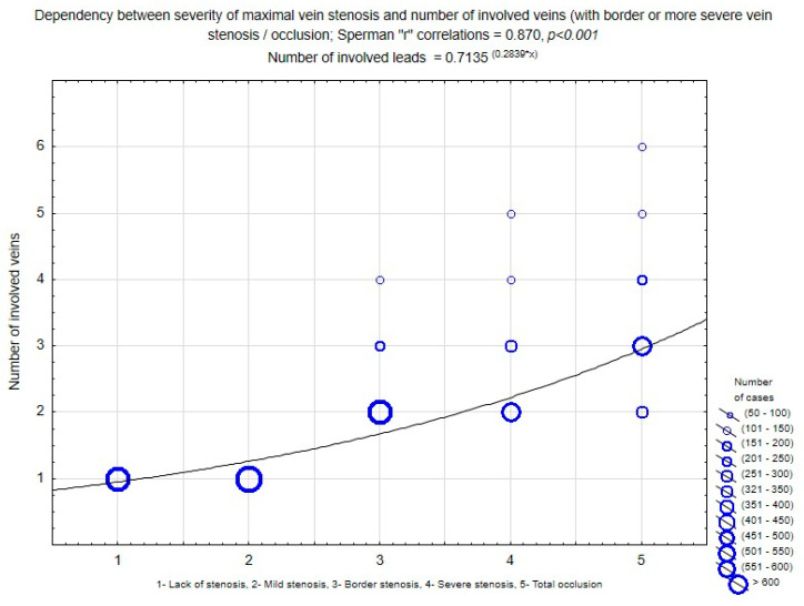
Background and Objectives: The nature of multilevel lead-related venous stenosis/occlusion (MLVSO) and its influence on transvenous lead extraction (TLE) as well as long-term survival remains poorly understood. Materials and Methods: A total of 3002 venograms obtained before a TLE were analyzed to identify the risk factors for MLVSO, as well as the procedure effectiveness and long-term survival. Results: An older patient age at the first system implantation (OR = 1.015; p < 0.001), the number of leads in the heart (OR = 1.556; p < 0.001), the placement of the coronary sinus (CS) lead (OR = 1.270; p = 0.027), leads on both sides of the chest (OR = 7.203; p < 0.001), and a previous device upgrade or downgrade with lead abandonment (OR = 2.298; p < 0.001) were the strongest predictors of MLVSO. Conclusions: The presence of MLVSO predisposes patients with cardiac implantable electronic devices (CIED) to the development of infectious complications. Patients with multiple narrowed veins are likely to undergo longer and more complex procedures with complications, and the rates of clinical and procedural success are lower in this group. Long-term survival after a TLE is similar in patients with MLVSO and those without venous obstruction. MLVSO probably better depicts the severity of global venous obstruction than the degree of vein narrowing at only one point.
Keywords: complications; long-term survival; multilevel lead-related venous obstruction; risk factors; transvenous lead extraction.
Conflict of interest statement
The authors declare no conflicts of interest.
Figures

Similar articles
-
Chronic venous obstruction during cardiac device revision: Incidence, predictors, and efficacy of percutaneous techniques to overcome the stenosis.Heart Rhythm. 2020 Feb;17(2):258-264. doi: 10.1016/j.hrthm.2019.08.012. Epub 2019 Aug 10. Heart Rhythm. 2020. PMID: 31404660
-
The use of laser lead extraction sheath in the presence of supra-cardiac occlusion of the central veins for cardiac implantable electronic device lead upgrade or revision.PLoS One. 2021 May 14;16(5):e0251829. doi: 10.1371/journal.pone.0251829. eCollection 2021. PLoS One. 2021. PMID: 33989335 Free PMC article.
-
Risk Factors and Long-Term Survival of Octogenarians and Nonagenarians Undergoing Transvenous Lead Extraction Procedures.Gerontology. 2021;67(1):36-48. doi: 10.1159/000511358. Epub 2020 Nov 26. Gerontology. 2021. PMID: 33242867
-
Lead-Related Venous Obstruction in Patients With Implanted Cardiac Devices: JACC Review Topic of the Week.J Am Coll Cardiol. 2022 Jan 25;79(3):299-308. doi: 10.1016/j.jacc.2021.11.017. J Am Coll Cardiol. 2022. PMID: 35057916 Review.
-
Venous thrombosis and stenosis after implantation of pacemakers and defibrillators.J Interv Card Electrophysiol. 2005 Jun;13(1):9-19. doi: 10.1007/s10840-005-1140-1. J Interv Card Electrophysiol. 2005. PMID: 15976973 Review.
Cited by
-
New Diseases Related to Cardiac Implantable Electronic Devices (CIEDs): An Overview.J Clin Med. 2025 Feb 17;14(4):1322. doi: 10.3390/jcm14041322. J Clin Med. 2025. PMID: 40004852 Free PMC article. Review.
References
-
- Lickfett L., Bitzen A., Arepally A., Nasir K., Wolpert C., Jeong K.M., Krause U., Schimpf R., Lewalter T., Calkins H., et al. Incidence of venous obstruction following insertion of an implantable cardioverter defibrillator. A study of systematic contrast venography on patients presenting for their first elective ICD generator replacement. Europace. 2004;6:25–31. doi: 10.1016/j.eupc.2003.09.001. - DOI - PubMed
MeSH terms
LinkOut - more resources
Full Text Sources
Medical
