

Isolation and Characterization of Yunnan Variants of the Pseudorabies Virus and Their Pathogenicity in Rats
- PMID: 38400009
- PMCID: PMC10891970
- DOI: 10.3390/v16020233
Isolation and Characterization of Yunnan Variants of the Pseudorabies Virus and Their Pathogenicity in Rats
Abstract
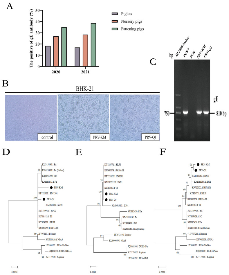
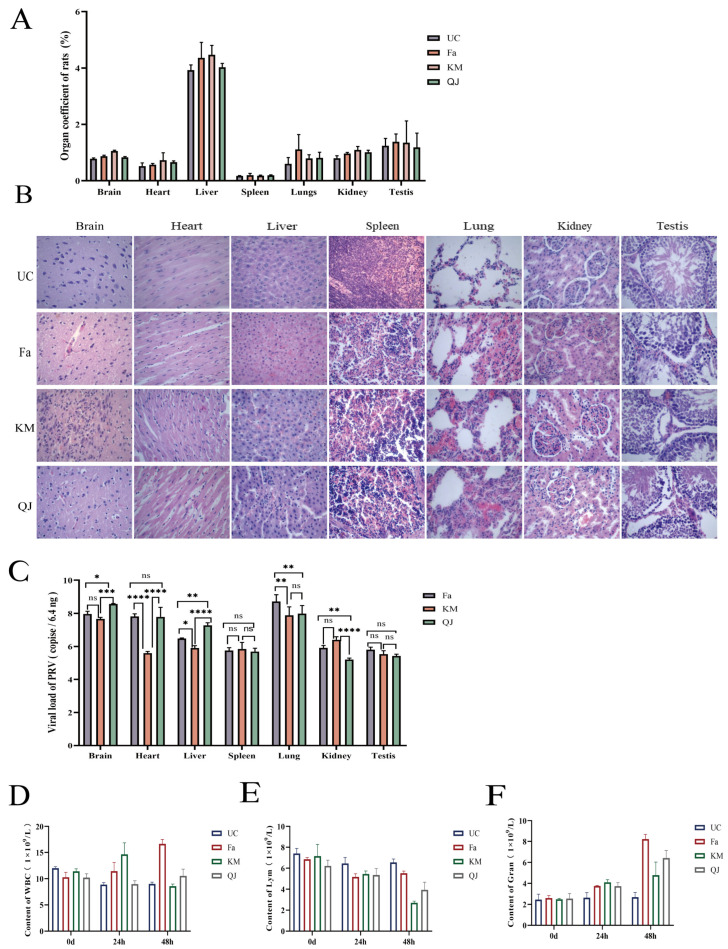
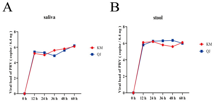
Porcine pseudorabies has long existed in China and is a serious threat to the Chinese farming industry. To understand the prevalence and genetic variation of the porcine pseudorabies virus (PRV) and its pathogenicity in Yunnan Province, China, we collected 560 serum samples across seven Yunnan Province regions from 2020 to 2021 and detected anti-gE antibodies in these samples. Sixty-one clinical tissue samples were also collected from pigs with suspected PRV that were vaccinated with Bartha-K61. PRV-gE antibodies were found in 29.6% (166/560) of the serum samples. The PRV positivity rate in clinical tissue samples was 13.1% (8/61). Two isolates, PRV-KM and PRV-QJ, were obtained. The identity of the gB, gD, and gE genes between these isolates and the Chinese mutants exceeded 99.5%. These isolates and the classical Fa strain were used to infect 4-week-old rats intranasally to assess their pathogenicity. All infected rats showed the typical clinical and pathological features of PRV two days post-infection. The viral loads in the organs differed significantly among the infected groups. Viruses were detected in the saliva and feces at 12 h. Significant dynamic changes in total white blood cell counts (WBC), lymphocyte counts (Lym), and neutrophil counts (Gran) occurred in the blood of the infected groups at 24 and 48 h. These results show that mutant PRV strains are prevalent in Bartha-K61-vaccinated pigs in Yunnan Province, China. Moreover, rats shed PRV in their saliva and feces during early infection, indicating the need for rodent control in combatting PRV infections in Yunnan Province, China.
Keywords: Yunnan; characterization; isolation; mutant strain; rat pathogenicity; swine pseudorabies.
Conflict of interest statement
The authors declare no conflicts of interest.
Figures
Similar articles
-
Pathogenicity and genomic characterization of a pseudorabies virus variant isolated from Bartha-K61-vaccinated swine population in China.Vet Microbiol. 2014 Nov 7;174(1-2):107-15. doi: 10.1016/j.vetmic.2014.09.003. Epub 2014 Sep 22. Vet Microbiol. 2014. PMID: 25293398
-
Emergence of a novel pathogenic recombinant virus from Bartha vaccine and variant pseudorabies virus in China.Transbound Emerg Dis. 2021 May;68(3):1454-1464. doi: 10.1111/tbed.13813. Epub 2020 Sep 8. Transbound Emerg Dis. 2021. PMID: 32857916
-
Pseudorabies virus variant in Bartha-K61-vaccinated pigs, China, 2012.Emerg Infect Dis. 2013 Nov;19(11):1749-55. doi: 10.3201/eid1911.130177. Emerg Infect Dis. 2013. PMID: 24188614 Free PMC article.
-
A Review of Pseudorabies Virus Variants: Genomics, Vaccination, Transmission, and Zoonotic Potential.Viruses. 2022 May 9;14(5):1003. doi: 10.3390/v14051003. Viruses. 2022. PMID: 35632745 Free PMC article. Review.
-
Pseudorabies Virus: From Pathogenesis to Prevention Strategies.Viruses. 2022 Jul 27;14(8):1638. doi: 10.3390/v14081638. Viruses. 2022. PMID: 36016260 Free PMC article. Review.
Cited by
-
Prevalence and Genetic Variation Investigation of the Pseudorabies Virus in Southwest China.Animals (Basel). 2024 Oct 28;14(21):3103. doi: 10.3390/ani14213103. Animals (Basel). 2024. PMID: 39518826 Free PMC article.
-
Alphaherpesvirus in Pets and Livestock.Microorganisms. 2025 Jan 4;13(1):82. doi: 10.3390/microorganisms13010082. Microorganisms. 2025. PMID: 39858850 Free PMC article. Review.
-
Molecular epidemiology and genetic characteristics of pseudorabies virus between 2021 and 2023 in Henan Province of China.J Vet Sci. 2025 Mar;26(2):e26. doi: 10.4142/jvs.24243. J Vet Sci. 2025. PMID: 40183912 Free PMC article.
-
Isolation and biological characterization of a variant pseudorabies virus strain from goats in Yunnan Province, China.Vet Res Commun. 2025 Mar 22;49(3):149. doi: 10.1007/s11259-025-10703-1. Vet Res Commun. 2025. PMID: 40119952
-
Astrocyte-derived MMP-9 is a key mediator of pseudorabies virus penetration of the blood-brain barrier and tight junction disruption.Vet Res. 2025 Apr 2;56(1):72. doi: 10.1186/s13567-025-01486-z. Vet Res. 2025. PMID: 40176142 Free PMC article.
References
-
- Hanson R.P. The history of pseudorabies in the United States. J. Am. Vet. Med. Assoc. 1954;124:259–261. - PubMed
MeSH terms
Substances
LinkOut - more resources
Full Text Sources
