. 2024 Feb 13;16(2):290.
doi: 10.3390/v16020290.

IE1 of Human Cytomegalovirus Inhibits Necroptotic Cell Death via Direct and Indirect Modulation of the Necrosome Complex

Affiliations

IE1 of Human Cytomegalovirus Inhibits Necroptotic Cell Death via Direct and Indirect Modulation of the Necrosome Complex

Anna Theresa Heusel et al. Viruses. .

Abstract

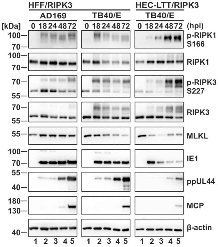
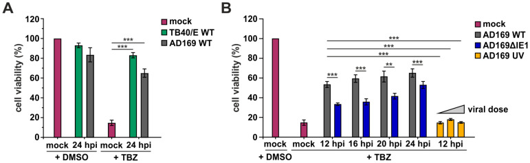
Programmed necrosis is an integral part of intrinsic immunity, serving to combat invading pathogens and restricting viral dissemination. The orchestration of necroptosis relies on a precise interplay within the necrosome complex, which consists of RIPK1, RIPK3 and MLKL. Human cytomegalovirus (HCMV) has been found to counteract the execution of necroptosis during infection. In this study, we identify the immediate-early 1 (IE1) protein as a key antagonist of necroptosis during HCMV infection. Infection data obtained in a necroptosis-sensitive cell culture system revealed a robust regulation of post-translational modifications (PTMs) of the necrosome complex as well as the importance of IE1 expression for an effective counteraction of necroptosis. Interaction analyses unveiled an association of IE1 and RIPK3, which occurs in an RHIM-domain independent manner. We propose that this interaction manipulates the PTMs of RIPK3 by promoting its ubiquitination. Furthermore, IE1 was found to exert an indirect activity by modulating the levels of MLKL via antagonizing its interferon-mediated upregulation. Overall, we claim that IE1 performs a broad modulation of innate immune signaling to impede the execution of necroptotic cell death, thereby generating a favorable environment for efficient viral replication.

Keywords: HCMV; IE1; MLKL; RIPK3; cytomegalovirus; innate immunity; interferon signaling; intrinsic immunity; necroptosis; necroptotic cell death.


Conflict of interest statement

The authors declare no conflicts of interest.

Figures

Figure 1
Figure 1
Generation of a necroptosis-sensitive cell culture model. (A) HFF and HEC-LTT cells were lentivirally transduced to stably express RIPK3. Successful integration of RIPK3 was monitored in Western blot analysis. (B) Expression of RIPK3 was analyzed by indirect immunofluorescence analysis. Cell nuclei were stained with DAPI. Scale bar, 100 µm. (C) The sensitivity of RIPK3 expressing cells to necroptosis was analyzed in a cell viability assay by monitoring intracellular ATP levels (CellTiter-Glo, Promega, Fitchburg, MA, USA). The cells were stimulated for 8 h with TBZ (TNFα (30 ng/mL), BV-6 (5 µM) and z-VAD-fmk (25 µM)). Green, DMSO-treated cells; grey, TBZ treated cells. Depicted values represent the means +/− SD derived from triplicates relative to control cells (%). (D) The modulation of the necrosome components after the indicated times of TBZ treatment was monitored in Western blot analysis. Β-actin served as internal loading control. Each experiment was performed three times in independent experiments and one representative experiment is shown. For statistical analysis a student’s t-test was performed (unpaired, two-tailed); *** p < 0.001.
Figure 2
Figure 2
Necrosome components are strongly modulated during HCMV infection. RIPK3 expressing HFF and HEC-LTT cells were infected for the indicated times and then harvested for Western blot analysis. HFF/RIPK3 cells were infected with AD169 (left panel) or TB40/E (middle panel) and HEC-LTT/RIPK3 with TB40/E (right panel). Infections were performed at an MOI of 5. Necrosome components and viral markers of infection were analyzed by Western blot experiments. Β-actin served as internal loading control. Three independent experiments were performed, and one representative experiment is shown.
Figure 3
Figure 3
HCMV-mediated rescue from necroptotic cell death begins already at the early stages of infection. HFF/RIPK3 cells were infected for the indicated times and subsequently treated with TBZ for 8 h or with DMSO as control. Cells were infected with TB40/E WT (A), AD169 WT (A,B) and AD169 ΔIE1 (B) at an MOI of 3. AD169 UV (B) was used at increasing viral doses (calculated MOIs of 6, 12 and 18). Necroptotic cell death was monitored by analyzing intracellular ATP levels using a cell viability assay (CellTiter-Glo, Promega, Fitchburg, MA, USA). Red, mock infected cells; green, TB40/E WT infected cells; grey, AD169 WT infected cells; blue, AD169ΔIE1 infected cells; yellow, AD169 UV infected cells. Depicted values (%) represent the mean +/− SD derived from triplicates relative to the control (mock, DMSO). Each experiment was performed two times in independent experiments and one representative experiment is shown. For statistical analysis a student’s t-test was performed (unpaired, two-tailed); ** p < 0.01, *** p < 0.001.
Figure 4
Figure 4
The immediate-early 1 (IE1) protein exhibits a strong anti-necroptotic activity by modulating the modification pattern of RIPK3. (A) HFF/RIPK3 cells with inducible IE1 expression (A,B) were treated with TBZ for the indicated times or with DMSO as control. Red, DMSO-treated cells; green, no IE1 expression; grey, IE1 expression. Depicted values represent the mean +/− SD derived from triplicates relative to the DMSO control (%). (B) Expression levels of p-RIPK3 S227, RIPK3 and MLKL were analyzed in presence or absence of IE1 after the indicated times of TBZ treatment by Western blot analysis. – IE1, no IE1 expression, + IE1, IE1 expression. (C) Expression plasmids of IE1 and RIPK3 were co-transfected in HEK293T cells, whereby increasing amounts of IE1 were used. The expression levels were monitored in Western blot analysis. (D) To confirm a ubiquitination of RIPK3 during necroptosis induction, HFF/RIPK3 cells were treated with DMSO or TBZ for 6 h +/− PYR-41 (inhibitor of ubiquitination, Selleck Chemicals LLC, Houston, TX, USA). (E) To confirm the ubiquitination of RIPK3 during HCMV infection, HFF/RIPK3 cells were infected with TB40/E at an MOI of 3 and at 24 hpi, PYR-41 was applied for 6 h. Protein levels of RIPK3 and IE1 were monitored in Western blot analysis. β-actin served as internal loading control. Each experiment was performed at least two times in independent experiments and always one representative experiment is shown. Fold changes of protein expression were determined according to signal intensities normalized to β-actin levels. For statistical analysis a student’s t-test was performed (unpaired, two-tailed); * p < 0.05, *** p < 0.001.
Figure 5
Figure 5
IE1 associates with the necrosome complex via binding RIPK3 and this interaction is promoted by distinct domains of IE1 and RIPK3. (A,C) HEK293T cells were co-transfected with expression plasmids for the indicated proteins and co-immunoprecipitation (Co-IP) was performed. FEN1-IE1 interaction served as positive control [37]. The IP of FLAG-tagged RIPK3, MLKL and FEN1 constructs was performed by using protein A-sepharose beads with immobilized anti-FLAG antibody. (B) HFF/control and HFF/RIPK3 were infected with TB40/E at an MOI of 1 for 4–18 h and subsequently IE1 was precipitated using protein A-sepharose beads with immobilized anti-IE1. (D) Schematic illustration of truncated variants of IE1 (upper panel) and RIPK3 (lower panel) which were used for fine-mapping of the interaction interface (E,F). NLS, nuclear localization sequence; core, globular core domain; STAT, binding domain of STAT proteins; CTD, chromatin tethering domain; RHIM, RIP homotypic interaction motif. (E,F) Fine-mapping of the interaction interface of IE1 and RIPK3 in HEK293T by transfecting expression plasmids encoding FLAG-tagged IE1 and RIPK3 variants and subsequent Co-IP analysis by using magnetic beads with immobilized anti-FLAG antibody. In (F), purified protein of IE1 was utilized. Each experiment was performed at least three times and one representative experiment is shown.
Figure 6
Figure 6
IE1 antagonizes the interferon-mediated upregulation of MLKL. HFF cells were treated with the indicated types of IFN (1000 U/mL) for 24 h. (A,C) Levels of necrosome components in the indicated HFF cells were analyzed in Western blot experiments. Β-actin served as internal loading control. Fold changes of protein expression were determined according to signal intensities normalized to β-actin levels. (C) − IE1, no IE1 expression; + IE1, IE1 expression; − IFN-β, no IFN-β stimulation; + IFN-β, IFN-β stimulation. (B,D) Modulation of MLKL transcription upon IFN-stimulation was analyzed by isolating total RNA and performing SYBR qPCR analysis in the indicated HFF cells. Depicted values represent the mean +/− SD derived from triplicates relative to untreated HFF/control cells and normalized to levels of the housekeeping gene GAPDH. Green, no IFN-β stimulation; grey, IFN-β stimulation. For statistical analysis a student’s t-test was performed with ΔCq-values (unpaired, two-tailed); ** p < 0.01, *** p < 0.001. Each experiment was performed three times in independent experiments and one representative experiment is shown.
Figure 7
Figure 7
IE1 modulates the activation of innate immune signaling to circumvent necroptosis. HFF/RIPK3 cells were infected with WT and ΔIE1 HCMV (both TB40/E) for 24 h at an MOI of 3. Subsequently, total RNA was isolated and necrosis/necroptosis-related profiling was performed by using the RT² Profiler Necrosis PCR array (Qiagen, Düsseldorf, Germany). Values above the dashed line (at Y = 1) indicate an upregulation and values below the dashed line indicate a downregulation during ΔIE1 infection compared to WT infection. Genes illustrated in pink are described as ISGs [48]. Depicted values represent the mean +/− SD derived from three repetitions, ΔIE1 relative to WT infected HFFs. For normalization the mean Cq-value of the housekeeping genes ACTB, B2M, HPRT1 and RPLP0 was utilized. For statistical analysis a multicomparison t-test was performed with ΔCq-values (unpaired, two-tailed); * p < 0.05, ** p < 0.01, *** p < 0.001.

Similar articles

Cited by

References

    1. Degterev A., Huang Z., Boyce M., Li Y., Jagtap P., Mizushima N., Cuny G.D., Mitchison T.J., Moskowitz M.A., Yuan J. Chemical inhibitor of nonapoptotic cell death with therapeutic potential for ischemic brain injury. Nat. Chem. Biol. 2005;1:112–119. doi: 10.1038/nchembio711. - DOI - PubMed
    1. Kerr J.F., Wyllie A.H., Currie A.R. Apoptosis: A basic biological phenomenon with wide-ranging implications in tissue kinetics. Br. J. Cancer. 1972;26:239–257. doi: 10.1038/bjc.1972.33. - DOI - PMC - PubMed
    1. Wu W., Liu P., Li J. Necroptosis: An emerging form of programmed cell death. Crit. Rev. Oncol. Hematol. 2012;82:249–258. doi: 10.1016/j.critrevonc.2011.08.004. - DOI - PubMed
    1. He S., Liang Y., Shao F., Wang X. Toll-like receptors activate programmed necrosis in macrophages through a receptor-interacting kinase-3-mediated pathway. Proc. Natl. Acad. Sci. USA. 2011;108:20054–20059. doi: 10.1073/pnas.1116302108. - DOI - PMC - PubMed
    1. Brault M., Olsen T.M., Martinez J., Stetson D.B., Oberst A. Intracellular Nucleic Acid Sensing Triggers Necroptosis through Synergistic Type I IFN and TNF Signaling. J. Immunol. 2018;200:2748–2756. doi: 10.4049/jimmunol.1701492. - DOI - PMC - PubMed

MeSH terms

Substances

LinkOut - more resources