. 2024 Feb 15;16(2):297.
doi: 10.3390/v16020297.

Validation of Candidate Host Cell Entry Factors for Bovine Herpes Virus Type-1 Based on a Genome-Wide CRISPR Knockout Screen

Affiliations

Validation of Candidate Host Cell Entry Factors for Bovine Herpes Virus Type-1 Based on a Genome-Wide CRISPR Knockout Screen

Wenfang Spring Tan et al. Viruses. .

Abstract

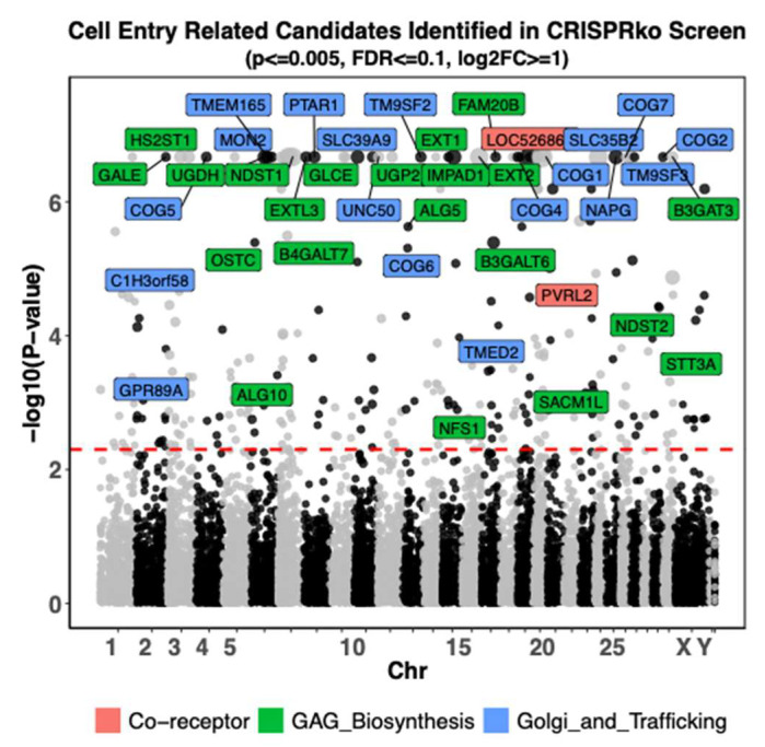
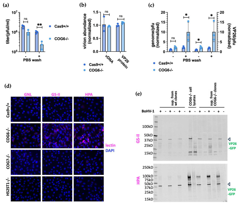
To identify host factors that affect Bovine Herpes Virus Type 1 (BoHV-1) infection we previously applied a genome wide CRISPR knockout screen targeting all bovine protein coding genes. By doing so we compiled a list of both pro-viral and anti-viral proteins involved in BoHV-1 replication. Here we provide further analysis of those that are potentially involved in viral entry into the host cell. We first generated single cell knockout clones deficient in some of the candidate genes for validation. We provide evidence that Polio Virus Receptor-related protein (PVRL2) serves as a receptor for BoHV-1, mediating more efficient entry than the previously identified Polio Virus Receptor (PVR). By knocking out two enzymes that catalyze HSPG chain elongation, HST2ST1 and GLCE, we further demonstrate the significance of HSPG in BoHV-1 entry. Another intriguing cluster of candidate genes, COG1, COG2 and COG4-7 encode six subunits of the Conserved Oligomeric Golgi (COG) complex. MDBK cells lacking COG6 produced fewer but bigger plaques compared to control cells, suggesting more efficient release of newly produced virions from these COG6 knockout cells, due to impaired HSPG biosynthesis. We further observed that viruses produced by the COG6 knockout cells consist of protein(s) with reduced N-glycosylation, potentially explaining their lower infectivity. To facilitate candidate validation, we also detailed a one-step multiplex CRISPR interference (CRISPRi) system, an orthogonal method to KO that enables quick and simultaneous deployment of three CRISPRs for efficient gene inactivation. Using CRISPR3i, we verified eight candidates that have been implicated in the synthesis of surface heparan sulfate proteoglycans (HSPGs). In summary, our experiments confirmed the two receptors PVR and PVRL2 for BoHV-1 entry into the host cell and other factors that affect this process, likely through the direct or indirect roles they play during HSPG synthesis and glycosylation of viral proteins.

Keywords: BoHV-1; COG complex; CRISPR/Cas9; Golgi apparatus; cell entry; heparan Sulfate; receptors.


Conflict of interest statement

The authors declare no conflict of interest. The funders had no role in the design of the study; in the collection, analyses, or interpretation of data; in the writing of the manuscript; or in the decision to publish the results.

Figures

Figure 1
Figure 1
Genome-wide CRISPR knockout screen identifies pro-viral genes associated with viral entry into the cell. Guide RNA copy number changes obtained from the CRISPRko screen by comparing GFP Negative cells to GFP High cells [49] were plotted. Each dot represents one of the 21,216 protein coding genes targeted by the btCRISPRko.v1 library, with its genomic location plotted against the x-axis and −log10 (p-value) based on MAGeCK76 analysis against the y-axis; the sizes of dots represent -log2FoldChange values. Pro-viral candidate genes that are most likely to affect BoHV-1 cell entry with p-value ≤ 0.005, False Discovery Rate (FDR) ≤ 0.1, and log2 fold changes (log2FC) ≥ 1 as statistical cutoff are highlighted; they are color coded based on the pathways they might impact. The red line represents p-value threshold as 0.005 or −log10(p-value) = 2.3. Chr: short for chromosome, due to space constraint, some chromosome numbers were omitted.
Figure 2
Figure 2
Loss of cell surface receptors impair BoHV-1 replication in MDBK cells. (a). Plaque assay results in four clones with PVR (a.k.a LOC526865) KO compared to Cas9+/+ control cells. (b). Plaque assay results in four clones with PVRL2-/- compared to Cas9+/+ controls cells. (c). Plaque assay results in four clones with PVRL2 and PVR double KO (dKO) compared to Cas9+/+ controls cells. All plaque assays shown in (ac) were done with at least two biological repeats (n ≥ 2), while plaque sizes were averages measured from at least 40 plaques and then normalized to those from Cas9+/+ cells. (d). BoHV-1 VP26-GFP protein growth curves in Cas9+/+, PVR-/-, PVRL2-/-, and PVR/PVRL2 dKO cells infected at MOI = 3 or 0.1 measured as fluorescence intensity (n = 4) throughout a 72-h infection. (e). Titers of total viruses harvested from non-KO cells (Cas9+/+ or WT cells) and PVR-/-; PVRL2-/- dKO cells at 16 h.p.i.(n = 2 with three clones each). More details described in 2.8. Briefly, ice cold virus inoculum was added at MOI = 1 to each cell line and the plates were incubated on ice. 1 h later (a.k.a. 0.h.p.i.), the inoculum was taken off and the cells were washed with ice cold PBS thrice before being returned to the 37 °C incubator. 3 h later, cells were washed again with an acid buffer (pH = 3) or PBS for 1 min and the plates were returned to the incubator till harvest. All analyses based on pairwise t-tests with ns: not significant with p > 0.05; *: p ≤ 0.05; **: p ≤ 0.01; ***: p ≤ 0.001; error bars represent +/- 1 SD.
Figure 3
Figure 3
Many candidates catalyze cell surface Heparan Sulfate proteoglycan synthesis. (a). Genes known to be involved in each step Heparan Sulfate (HS) biosynthesis and chain extension from the core protein (red squiggle); those highlighted in blue were identified by the screen. This diagram is modeled after Figure 2 in Chen et al. [55] with the pathway to chondroitin sulfate (CS)/dermatan sulfate (DS) synthesis simplified. Abbreviations for units within the HS chain: Xyl, xylose residue; Gal, galactose residue; GlcNAc, 2-deoxy-2-acetamido-α-D-glucopyranosyl; GlcA, β-D-glucuronic acid; IdoA, α-L-iduronic acid. (b). Cells were treated with specified concentrations of Heparin or Chondroiten Sulfate prior to infection by BoHV-1. Total viral samples were harvested and titrated on wt MDBKs cells (n = 3 for HS blocking assay and n = 2 for CS blocking). (c). Plaque assay results from four GLCE-/- clones, A1, A7, B2 and B8 compared to Cas9+/+ control cells (n ≥ 6). Plaque size measurements were taken from at least 50 plaques for each clone and then normalized to those from Cas9+/+ cells. (d). Plaque assay results from four HST2ST1-/- clones, A5, C1, C2 and C4 compared to Cas9+/+ control cells (n ≥ 5). Plaque size measurements were taken from at least 89 plaques for each clone and normalized to Cas9+/+ cells. (e). Titers of total viruses grown in non-KO cells and HST2ST1-/- KO cells (MOI = 1) harvested at 16 h.p.i. The experiment was conducted the same as described in Figure 2e and Section 2.8. All analyses based on pairwise t-tests with ns: not significant with p > 0.05; **: p ≤ 0.01; ***: p ≤ 0.001; error bars represent +/− 1 SD.
Figure 4
Figure 4
The loss of COG6 and COG7 affects BoHV-1 replication in MDBK cells. (a). Plaque assay results from five COG6-/- KO clones compared to Cas9+/+ control cells with measured virus titers (left) and plaque sizes (right) shown. (b). Plaque assay results from four COG7-/- KO clones compared to Cas9+/+ control cells with measured virus titer(left) and plaque sizes shown (right). Statistics for A and B as follows: all plaque assays were conducted at least three times for each clone (n ≥ 3) with plaque sizes averaged from ≥ 65 plaques for each KO clone and then normalized to that from Cas9+/+ cells. ***: p < 0.005; *: p < 0.05; n.s.: not significant based on one-way ANOVA followed by multiple comparisons between Cas9+/+ and Cog7-/- clones, error bars represent +/− 1 SD. (c). VP26-GFP fluorescence growth curve in Cas9+/+ and COG KO cells infected with GFP tagged BoHV-1 at MOI = 3 or 0.1. (d). Plaque assay results by titrating virus harvested from COG6-/- KO or Cas9+/+ cells infected with BoHV-1 at MOI = 0.1 on wt MDBK cells, supernatant and cell pellet samples were collected at 0,24,48,72 h.p.i. Two-way ANOVA followed by Šídák’s multiple comparisons test was performed to compare the two cell lines (n = 3, ****: p < 0.0001). The error bars (+/− 1 SD) are too small to be displayed. (e). Titers of total viruses grown in non-KO cells and COG6 or COG7 KO cells (MOI = 1) harvested at 16 h.p.i. The experiment was conducted the same as described in Figure 2e and Section 2.8. All analyses except the time course experiment (d) based on pairwise t-tests with ns: not significant with p > 0.05; *: p ≤ 0.05; ***: p ≤ 0.001; ****: p ≤ 0.0001; error bars represent +/- 1 SD.
Figure 5
Figure 5
The loss of COG6 lead to the production of less infectious viruses possibly due to reduced glycosylation of viral protein(s). (a). Titers of supernatant viruses harvested (at 48 h.p.i. with MOI = 0.1) from COG6-/- or Cas9+/+ cells plaqued on wild type MDBK cells. For one set of the plaque assays, the inoculums were taken off one hour after addition of the viruses and the MDBK monolayers were washed three times with PBS. For the other set, the inoculums remained. (b). Quantification of viral particles in the samples based on the viral genome copy number (vDNA, measured by qPCR) and the VP26 protein abundance (measured by densitometry of western blot results shown in (e)). (c). Normalized genome/pfu ratios and VP26 protein/pfu ratios of these viruses. (d). Lectin staining results of the COG6-/-, COG7-/- and HST2ST1-/- clones and Cas9+/+ cells using GNL, GS-II and HPA dyes. The staining was visualized by fluorescent microscopy with positive lectin stain shown in pink while DAPI counterstain of the nuclei in blue. Representative results from three independent clones shown. (e). Lectin staining (GS-II and HPA) coupled with western blotting (against VP26-GFP) of concentrated viruses from (a). Concentrated supernatants from MOCK infected clones and COG6-/- cell lysates served as controls. Results shown are from three independent clones for COG6-/- and Cas9+/+ (wt). All analyses are based on pairwise t-tests with ns: not significant with p > 0.05; *: p ≤ 0.05; **: p ≤ 0.01; error bars represent +/− 1 SD.
Figure 6
Figure 6
CRISPRi mediates efficient KD of gene expression in MDBK cells. (a). Schematic representation of the CRISPRi system applied in this study, based on the module developed by Andrea Califano et al. (See Materials and Methods). (b). Doxycycline inducible expression of the dCas9-KRAB-MeCP2 module from a stably integrated expression cassette at the cow rosa26 locus. Green heptagram represents doxycycline, purple squares are rTTA expressed from the downstream expression cassette, used to enhance doxycycline-driven induction. (c). Targeting strategy to knock-in the dCas9-KRAB-MeCP2 cassette into rosa26 using TAL1.6 stimulated HDR) [49] and Hygromycin selection, prior to dilutional cloning and genotyping by PCR using primer set F + R (red arrows) binding outside the homology arms. wt: wild type; tg: targeted. (d). Representative genotyping results by PCR from a homozygous targeted clone (+/+), and two heterozygotes (+/wt), black arrows point to amplicons from the targeted allele (top, 12 kb PCR product) and wt allele (bottom, 4 kb PCR product). (e). Knockdown efficiency of Doxycycline induced CRISPRi under six Doxycycline concentrations across five genes represented as mRNA level measured by reverse transcription and qPCR at 48 h post induction. (f). Relative expression levels of five genes in dCas9+/+ MDBKs each targeted by three CRISPRi guides (g1, g2, g3) compared to cells transfected with non-targeting CRISPRs (NC). These results are based on RT-qPCR results from a single experiment with three technical qPCR repeats, error bars represent +/- 1 standard error of the mean.
Figure 7
Figure 7
Multiplex CRISPRi against pro-viral candidate genes results in reduced viral titers. (a). CRISPR3i against a host gene in action by binding immediately downstream of TSS in tandem for synergistic gene repression. (b). The one-step cloning strategy to synthesize PiggyBac transposon vectors carrying three sgRNA expression cassettes in tandem to implement CRISPR3i by PB transposition. (c). Plaque formation assay results in cells with CRISPR3i expression intended to interfere with transcription of pro-viral genes involved in HS biosynthesis and other functions, as identified by the CRISPRko screen; CTRL represents virus titer from cells expressing three non-targeting sgRNAs (n >= 3). Results were ordered by p-value. ****: p < 0.0001; ***: p < 0.005; **: p < 0.01; *: p < 0.05; n.s. not significant with p > 0.05 based on one-way ANOVA followed by multiple comparisons between CTRL and CRISPR3i cells, error bars represent +/− 1 SD.

Similar articles

References

    1. Jones C., Chowdhury S. A review of the biology of bovine herpesvirus type 1 (BHV-1), its role as a cofactor in the bovine respiratory disease complex and development of improved vaccines. Anim. Health Res. Rev. 1996;8:187–205. doi: 10.1017/S146625230700134X. - DOI - PubMed
    1. Babiuk L.A., van Drunen Littel-van den Hurk S., Tikoo S.K. Immunology of bovine herpesvirus 1 infection. Veter. Microbiol. 1996;53:31–42. doi: 10.1016/S0378-1135(96)01232-1. - DOI - PubMed
    1. Srikumaran S., Kelling C.L., Ambagala A. Immune evasion by pathogens of bovine respiratory disease complex. Anim. Health Res. Rev. 1996;8:215–229. doi: 10.1017/S1466252307001326. - DOI - PubMed
    1. Pastenkos G., Lee B., Pritchard S.M., Nicola A.V. Bovine Herpesvirus 1 Entry by a Low-pH Endosomal Pathway. J. Virol. 2018;92:e00839-18. doi: 10.1128/JVI.00839-18. - DOI - PMC - PubMed
    1. Liang X.P., Babiuk L.A., van Drunen Littel-van den Hurk S., Fitzpatrick D.R., Zamb T.J. Bovine herpesvirus 1 attachment to permissive cells is mediated by its major glycoproteins gI, gIII, and gIV. J. Virol. 1991;65:1124–1132. doi: 10.1128/jvi.65.3.1124-1132.1991. - DOI - PMC - PubMed