. 2024 Feb 2;12(2):157.
doi: 10.3390/vaccines12020157.

Development of NP-Based Universal Vaccine for Influenza A Viruses

Affiliations

Development of NP-Based Universal Vaccine for Influenza A Viruses

Ekramy E Sayedahmed et al. Vaccines (Basel). .

Abstract

The nucleoprotein (NP) is a vital target for the heterosubtypic immunity of CD8+ cytotoxic T lymphocytes (CTLs) due to its conservation among influenza virus subtypes. To further enhance the T cell immunity of NP, autophagy-inducing peptide C5 (AIP-C5) from the CFP10 protein of Mycobacterium tuberculosis was used. Mice were immunized intranasally (i.n.) with human adenoviral vectors, HAd-C5-NP(H7N9) or HAd-NP(H7N9), expressing NP of an H7N9 influenza virus with or without the AIP-C5, respectively. Both vaccines developed similar levels of NP-specific systemic and mucosal antibody titers; however, there was a significantly higher number of NP-specific CD8 T cells secreting interferon-gamma (IFN-γ) in the HAd-C5-NP(H7N9) group than in the HAd-NP(H7N9) group. The HAd-C5-NP(H7N9) vaccine provided better protection following the challenge with A/Puerto Rico/8/1934(H1N1), A/Hong Kong/1/68(H3N2), A/chukkar/MN/14951-7/1998(H5N2), A/goose/Nebraska/17097/2011(H7N9), or A/Hong Kong/1073/1999(H9N2) influenza viruses compared to the HAd-NP(H7N9) group. The autophagy transcriptomic gene analysis of the HAd-C5-NP(H7N9) group revealed the upregulation of some genes involved in the positive regulation of the autophagy process. The results support further exploring the use of NP and AIP-C5 for developing a universal influenza vaccine for pandemic preparedness.

Keywords: adenoviral vector; autophagy; autophagy-inducing peptide; influenza vaccine; nucleoprotein; universal influenza vaccine.


Conflict of interest statement

The authors declare no conflicts of interest.

Figures
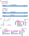
Figure 1
Figure 1
(A) Diagrammatic image of HAd−C5−NP(H7N9), HAd−NP(H7N9), and HAd-ΔE1E3 genomic representation. The cytomegalovirus (CMV) promoter and the bovine growth hormone (BGH) polyadenylation signal are flanking the NP(H7N9) or C5-NP(H7N9) genes. The drawings are not to scale. LITR, left inverted terminal repeat; RITR, right inverted terminal repeat; ΔE1, deletion of E1 region; ΔE3, deletion of E3 region; C5, C5-AIP; NP, nucleoprotein. (B) Immunoblot confirming expression of NP(H7N9) or C5-NP(H7N9) in HAd-NP(H7N9)- or HAd-C5-NP(H7N9)-infected 293 cells, respectively. Mock or HAd-ΔE1E3 infected cell extracts were used as a negative control. The molecular weight marker is shown on the left. Outlines of the one-dose (C) or the two-dose (D) animal inoculation study.
Figure 2
Figure 2
Immunogenicity of HAd−NP(H7N9) or HAd−C5−NP(H7N9) in inoculated mice. BALB/c mice six to eight weeks old (5 animals/group) were vaccinated once intranasally (i.n.), as described in the material and methods section. After vaccination for four weeks, blood samples were collected and used to monitor the development of NP-specific IgG (A), IgG1 (B), IgG2a (C), and IgA (D) antibody responses by ELISA. Lung washes were also collected to monitor the development of mucosal NP-specific IgG (E), IgG1 (F), IgG2a (G), and IgA (H) antibody responses by ELISA. The data are displayed as the mean ± standard deviation (SD) of the optical density (OD). Enhancement in the number of NP-specific IFN-γ-secreting CD8 T cells following immunization with HAd-C5-NP(H7N9) was monitored at 4 weeks post-vaccination in the spleen (I), mediastinal lymph node (LN) (J), and lung mononuclear (MN) Cells (K) by enumerating NP-specific IFN-γ-secreting CD8 T cells by ELISpot using the NP-147 peptide. ns, non-significant at p > 0.05; *, significant at p < 0.05; **, significant at p < 0.01; ***, significant at p < 0.001; and ****, significant at p < 0.0001.
Figure 3
Figure 3
Protection efficacy of single i.n. vaccination of mice with Had-NP(H7N9) or Had-C5-NP(H7N9). At 4 weeks post-inoculation, immunized animal groups were challenged with 2 lethal doses of 50 (LD50) of A/Puerto Rico/8/1934(H1N1) (A,B) or 5 LD50 of A/Hong Kong/1/68(H3N2) (C,D). (A,C) Morbidity and (B,D) mortality after challenge were monitored. (EG). Groups were challenged with 100 mouse infectious dose 50 (MID50) of A/chukkar/MN/14951-7/1998(H5N2) (E), A/goose/Nebraska/17097/2011(H7N9) (F), or A/Hong Kong/1073/1999(H9N2) (G) influenza virus, and at 3 days post-challenge, the lungs were collected for virus titers. The data are shown as mean Log10 tissue culture infectious dose 50 (TCID50) or egg infectious dose 50 (EID50), and the detection limit was 0.5 Log10 TCID50 or EID50 per ml. ns, non-significant at p > 0.05; *, significant at p < 0.05; **, significant at p < 0.01; ***, significant at p < 0.001; and ****, significant at p < 0.0001.
Figure 4
Figure 4
Immunogenicity of Had-NP(H7N9) or Had-C5-NP(H7N9) two-dose regimen inoculation. BALB/c mice six to eight weeks old (5 animals/group) were immunized twice intranasally (i.n.), as mentioned in the material and methods section. Three weeks post-boost, blood samples were collected and used to monitor the development of NP-specific IgG (A), IgG1 (B), IgG2a (C), and IgA (D) antibody responses by ELISA. Three weeks post-boost, lung washes were also collected to monitor the development of mucosal NP-specific IgG (E), IgG1 (F), IgG2a (G), and IgA (H) antibody responses by ELISA. ELISA results are the optical density (OD) readings as mean ± standard deviation (SD). Enhancement in the number of NP-specific IFN-γ-secreting CD8 T cells following immunization with HAd-C5-NP(H7N9) was monitored at 3 weeks post-booster in the spleen (I), mediastinal lymph node (LN) (J), and lung mononuclear (MN) cells (K) by enumerating NP-specific IFN-γ-secreting CD8 T cells by ELISpot using the NP-147 peptide. ns, non-significant at p > 0.05; **, significant at p < 0.01; ***, significant at p < 0.001; and ****, significant at p < 0.0001.
Figure 5
Figure 5
Protection of Had-NP(H7N9) or Had-C5-NP(H7N9) two-dose regimen. At 3 weeks post-booster, immunized animal groups were challenged with 2 lethal doses of 50 (LD50) of A/Puerto Rico/8/1934(H1N1) (A,B) or 5 LD50 of A/Hong Kong/1/68(H3N2) (C,D). (A,C) Morbidity and (B,D) mortality after challenge were monitored. (EG). Groups were challenged with 100 mouse infectious dose 50 (MID50) of A/chukkar/MN/14951-7/1998(H5N2) (E), A/goose/Nebraska/17097/2011(H7N9) (F), or A/Hong Kong/1073/1999(H9N2) (G) influenza virus, and the lungs were collected 3 days post-challenge and lung viral titers were determined. The data are shown as mean Log10 tissue culture infectious dose 50 (TCID50) or egg infectious dose 50 (EID50), and the detection limit was 0.5 Log10 TCID50 or EID50 per ml. ns, non-significant at p > 0.05; and ****, significant at p < 0.0001.
Figure 6
Figure 6
Histopathology of HAd−NP(H7N9)- or HAd−C5−NP(H7N9)-immunized mice lung tissues. (A) BALB/c mice (3 animals/group) were mock-immunized (PBS) or immunized intranasally (i.n.) with 108 plaque-forming units (PFU) of HAd-∆E1E3, HAd-NP(H7N9), or HAd-C5-NP(H7N9). Animals were euthanized at 0.25-, 0.5-, 1-, 2-, 4-, and 8-day post-immunization, and the lung tissue samples were collected and processed for histopathology. Representative pictures of each group on days 1, 2, 4, and 8 post-immunization are shown (H&E, 200X). (B) The lung tissue histopathological scores from HAd-NP(H7N9)- or HAd-C5-NP(H7N9)-immunized mice. Lung tissue sections were blindly analyzed by a board-certified veterinary pathologist. ns, non-significant at p > 0.05; and *, significant at p < 0.05.
Figure 7
Figure 7
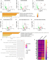
AIP-C5-mediated upregulation of genes involved in autophagy. BALB/c mice (3 animals/group) were mock-inoculated with PBS or inoculated i.n. once with 1 × 10⁸ PFU of Had-NP(H7N9), Had-C5-NP(H7N9), or HAd-ΔE1E3. The animals were euthanized at 24 h post-inoculation, and the lungs were collected for total RNA extraction. The RT2 mouse autophagy PCR array was used to analyze the differentially expressed genes in all study groups. (A) The differentially expressed genes between the HAd-∆E1E3 and the PBS groups are presented as a volcano plot. (B) The differentially expressed genes between the HAd-NP(H7N9) and the PBS groups are presented as a volcano plot. (C) The differentially expressed genes between the HAd-C5-NP(H7N9) and the PBS groups are presented as a volcano plot. (D) A bar graph showing the biological pathways of the significantly upregulated gene list input of the HAd-C5-NP(H7N9) group over the PBS group, colored by the p-values, is presented using the Metascape web analysis tool. (E) The differentially expressed genes between the HAd-NP(H7N9) and the HAd-∆E1E3 groups are presented as a volcano plot. (F) The differentially expressed genes between the HAd-C5-NP(H7N9) and the HAd-∆E1E3 groups are presented as a volcano plot. (G) The differentially expressed genes between the HAd-C5-NP(H7N9) and the HAd-NP(H7N9) groups are presented as a volcano plot. (H) A bar graph showing the biological pathways of the significantly upregulated gene list input of the HAd-C5-NP(H7N9) group over the HAd-∆E1E3 group, colored by the p-values, is presented using the Metascape web analysis tool. (I) A dot blot chart of the gene ontology enrichment analysis for the biological pathways involved in the upregulated gene list of the HAd-C5-NP(H7N9) group using ShinyGO v0.76 is shown. (J) A heat map of the differentially expressed genes of the HAd-NP(H7N9) group and HAd-C5-NP(H7N9) group compared to the HAd-∆E1E3 group.

Similar articles

Cited by

References

    1. Krammer F., Smith G.J.D., Fouchier R.A.M., Peiris M., Kedzierska K., Doherty P.C., Palese P., Shaw M.L., Treanor J., Webster R.G., et al. Influenza. Nat. Rev. Dis. Primers. 2018;4:3. doi: 10.1038/s41572-018-0002-y. - DOI - PMC - PubMed
    1. WHO Influenza (Seasonal) [(accessed on 26 January 2024)]. Available online: https://www.who.int/news-room/fact-sheets/detail/influenza-(seasonal)
    1. Sutton T.C. The Pandemic Threat of Emerging H5 and H7 Avian Influenza Viruses. Viruses. 2018;10:461. doi: 10.3390/v10090461. - DOI - PMC - PubMed
    1. WHO Influenza (Avian and Other Zoonotic) [(accessed on 21 September 2022)]. Available online: https://www.who.int/news-room/fact-sheets/detail/influenza-(avian-and-ot...
    1. More S., Bicout D., Bøtner A., Butterworth A., Calistri P., Depner K., Edwards S., Garin-Bastuji B., Good M., Gortázar Schmidt C., et al. Avian influenza. Efsa J. 2017;15:e04991. doi: 10.2903/j.efsa.2017.4991. - DOI - PMC - PubMed