. 2024 Apr 25;67(8):6081-6098.
doi: 10.1021/acs.jmedchem.3c01036. Epub 2024 Feb 24.

Paraptotic Cell Death as an Unprecedented Mode of Action Observed for New Bipyridine-Silver(I) Compounds Bearing Phosphane Coligands

Affiliations

Paraptotic Cell Death as an Unprecedented Mode of Action Observed for New Bipyridine-Silver(I) Compounds Bearing Phosphane Coligands

Ricardo G Teixeira et al. J Med Chem. .

Abstract

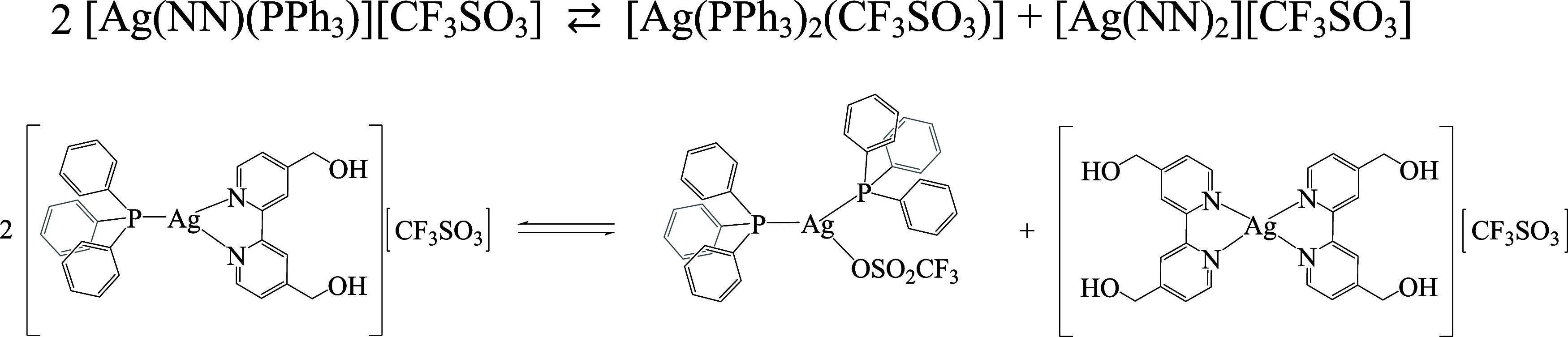
In this work, we investigated the anticancer activity of several novel silver(I) 2,2'-bipyridine complexes containing either triphenylphosphane (PPh3) or 1,2-bis(diphenylphosphino)ethane (dppe) ligands. All compounds were characterized by diverse analytical methods including ESI-MS spectrometry; NMR, UV-vis, and FTIR spectroscopies; and elemental analysis. Moreover, several compounds were also studied by X-ray single-crystal diffraction. Subsequently, the compounds were investigated for their anticancer activity against drug-resistant and -sensitive cancer cells. Noteworthily, neither carboplatin and oxaliplatin resistance nor p53 deletion impacted on their anticancer efficacy. MES-OV cells displayed exceptional hypersensitivity to the dppe-containing drugs. This effect was not based on thioredoxin reductase inhibition, enhanced drug uptake, or apoptosis induction. In contrast, dppe silver drugs induced paraptosis, a novel recently described form of programmed cell death. Together with the good tumor specificity of this compound's class, this work suggests that dppe-containing silver complexes could be interesting drug candidates for the treatment of resistant ovarian cancer.


Conflict of interest statement

The authors declare no competing financial interest.

Figures

Figure 1
Figure 1
Chemical structures of compounds 17. All compounds were isolated as CF3SO3 salts.
Figure 2
Figure 2
31P{1H} NMR spectra (162 MHz, CDCl3) of the PPh3-containing compounds 2 (A) and 3 (B) at different temperatures.
Scheme 1
Scheme 1. Depictive Reaction Equilibrium Detected for Complex 3 to Yield the Additional AgP2 Species in the Solution (CDCl3) Observed by NMR and MS
Figure 3
Figure 3
31P{1H} NMR spectra (162 MHz, CDCl3) of the dppe-containing compounds 5 (A) and 6 (B) at different temperatures.
Figure 4
Figure 4
ORTEP plots (50% probability) and labeling schemes for compounds 1 (A), 2 (B), 3 (C), and 4 (D). Solvent molecules and disordered atoms are omitted for clarity.
Figure 5
Figure 5
DFT optimized structures of complexes 1 (top, left) and 4 (top, right). The optimized structure of the proposed complex formed by 4 at low temperatures by VT-NMR studies ([Ag2(NN)2(μ-dppe)2]2+, 4*) is shown on the bottom. Hydrogen atoms are omitted for clarity. Experimental X-ray values are given in parentheses. Distances (bold) are given in angstroms, and angles (italics) are given in degrees.
Figure 6
Figure 6
Intracellular silver levels after treatment with 1, 4, or 7 (10 μM) in SK-OV-3 and MES-OV after 5 h were determined by ICP-MS. Results were normalized to the cell number, and values are given as mean ± SD of two independent experiments. Statistical significance was tested by two-way ANOVA and Sidak′s multiple comparison test (**p < 0.01; *p < 0.05).
Figure 7
Figure 7
TrxR activity after incubation with different concentrations of the selected compounds (IC50 [μM] ± SD) 1 (▲): 0.093 ± 0.014; 4 (■): 0.101 ± 0.009; 7 (●): 1.021 ± 0.192.
Figure 8
Figure 8
Apoptosis induction by the novel silver drugs in SK-OV-3 and MES-OV cells. (A) Cell death was determined by annexin V/propidium iodide (AV/PI) stain by flow cytometry after 24 h treatment with the indicated concentrations. Means ± SD were derived from three independent experiments. Significance to control was calculated by two-way ANOVA and Bonferroni’s multiple comparison test using the GraphPad Prism software (***p < 0.001, **p ≤ 0.01, *p ≤ 0.05). (B) Protein expression of PARP and cleaved PARP-1 as a late-phase apoptosis marker as well as γH2AX as a DNA damage marker was detected by Western blot after 24 h treatment with the tested silver compounds at the indicated concentrations.
Figure 9
Figure 9
Cytoplasmatic vacuoles indicative for paraptotic cell death induced by silver drug treatment. (A) Phase contrast images of MES-OV cells treated with the indicated drugs for 24 h (20× magnification, scale bar: 100 μm). (B) Percentage of vacuolated cells counted from phase-contrast microscopy images seen in panel A and Figure S42 after 6 and 24 h treatment. Values given are the mean ± standard deviation of three images with at least 30 cells in total. (C) Spinning disk confocal microscopy of ER-YFP-transfected SW480 cells treated with the indicated drugs and concentrations for 24 h. Representative pictures were taken in confocal mode, Z-stack, and max intensity projection (192× magnification and 60× objectives) of vesicles in ER-YFP-transfected SW480 (ER-YFP in green), DIC (differential interference contrast), and mitochondria (MitoTracker in red).
Figure 10
Figure 10
TEM analyses of MES-OV cells after 24 h treatment with the indicated drugs. Control cells exhibited a normal subcellular organization. In 1-treated samples small, single-membrane vacuoles (asterisks) and slightly thicker rough ER (black arrows) are indicated. Frequently, lysosomes and mitochondria exhibit degradation (asterisks). Upon therapy with 4, swollen rough ER (black arrows) and lysosomes with irregular lamellar structures were present (asterisk). Other cells exhibited stronger vacuolization (asterisks). In 7-treated samples, the cytoplasm was mostly devoid of organelles and filled with vacuoles (asterisks). Frequently, multilamellar bodies (white arrow) were found. Abbreviations: go = Golgi, ly = lysosome, mi = mitochondrium, nu = nucleus, black arrow = rough endoplasmatic reticulum, white arrow = multilamellar body, asterisk = single-membrane vacuole. All scalebars = 1 μm.
Figure 11
Figure 11
Impact of the MAPK inhibitor U0126 (5 and 10 μM) on the anticancer activity of the tested silver compounds. To evaluate the cells' viability, an MTT assay was performed after 72 h of combined drug treatment. The mean ± SD was derived from triplicates of one representative experiment out of three.
Figure 12
Figure 12
(A) Protein expression of ER-stress markers was detected by Western blot after 24 h treatment with the tested silver compounds at the indicated concentrations. (B) ROS production was measured by flow cytometry of DCF-DA fluorescence in MES-OV cells treated with indicated concentrations of the tested silver complexes after 5 and 24 h. Significance to control was calculated with one-way ANOVA and Dunnett’s multiple comparison test (****p < 0.0001, ***p < 0.0002, **p ≤ 0.002, *p ≤ 0.03, ns 0.12). (C) Changes in total, reduced and oxidized glutathione (GSH vs GSSG) are shown by fold increase to control of luminescence in MES-OV cells treated with the indicated drugs for 24 h. Significance to control was calculated with two-way ANOVA and Tukey’s multiple comparison test (****p < 0.0001, ***p < 0.0002, **p ≤ 0.002, *p ≤ 0.03, ns 0.12).

Similar articles

Cited by

References

    1. Ferlay J.; Soerjomataram I.; Dikshit R.; Eser S.; Mathers C.; Rebelo M.; Parkin D. M.; Forman D.; Bray F. Cancer Incidence and Mortality Worldwide: Sources, Methods and Major Patterns in GLOBOCAN 2012. Int. J. Cancer 2015, 136 (5), E359–E386. 10.1002/ijc.29210. - DOI - PubMed
    1. Reid B. M.; Permuth J. B.; Sellers T. A. Epidemiology of Ovarian Cancer: A Review. Cancer Biol. Med. 2017, 14 (1), 9–32. 10.20892/j.issn.2095-3941.2016.0084. - DOI - PMC - PubMed
    1. Pietragalla A.; Ciccarone F.; Nero C.; Scambia G.; Lorusso D.; Daniele G. Integration of PARP-Inhibitors in Ovarian Cancer Therapy. Explor. Target. Anti-tumor Ther. 2020, 1 (3), 171–182. 10.37349/etat.2020.00011. - DOI - PMC - PubMed
    1. Bonadio R. C.; Estevez-Diz M. D. P. Perspectives on PARP Inhibitor Combinations for Ovarian Cancer. Front. Oncol. 2021, 11 (December), 1–12. 10.3389/fonc.2021.754524. - DOI - PMC - PubMed
    1. O’Cearbhaill R. E. Using PARP Inhibitors in Advanced Ovarian Cancer Running Head: PARP Inhibitors in Ovarian Cancer. Oncology 2018, 32 (7), 339–343. - PMC - PubMed

Publication types

MeSH terms