. 2024 Apr 4;84(7):1290-1303.e7.
doi: 10.1016/j.molcel.2024.01.029. Epub 2024 Feb 23.

Bidirectional substrate shuttling between the 26S proteasome and the Cdc48 ATPase promotes protein degradation

Affiliations

Bidirectional substrate shuttling between the 26S proteasome and the Cdc48 ATPase promotes protein degradation

Hao Li et al. Mol Cell. .

Abstract

Most eukaryotic proteins are degraded by the 26S proteasome after modification with a polyubiquitin chain. Substrates lacking unstructured segments cannot be degraded directly and require prior unfolding by the Cdc48 ATPase (p97 or VCP in mammals) in complex with its ubiquitin-binding partner Ufd1-Npl4 (UN). Here, we use purified yeast components to reconstitute Cdc48-dependent degradation of well-folded model substrates by the proteasome. We show that a minimal system consists of the 26S proteasome, the Cdc48-UN ATPase complex, the proteasome cofactor Rad23, and the Cdc48 cofactors Ubx5 and Shp1. Rad23 and Ubx5 stimulate polyubiquitin binding to the 26S proteasome and the Cdc48-UN complex, respectively, allowing these machines to compete for substrates before and after their unfolding. Shp1 stimulates protein unfolding by the Cdc48-UN complex rather than substrate recruitment. Experiments in yeast cells confirm that many proteins undergo bidirectional substrate shuttling between the 26S proteasome and Cdc48 ATPase before being degraded.

Keywords: AAA ATPase; Cdc48; ERAD; p97; proteasome; protein degradation; protein unfolding; shuttling factor; ubiquitin.


Conflict of interest statement

Declaration of interests The authors declare no competing interests.

Figures
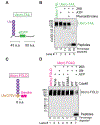
Figure 1.
Figure 1.. Cdc48-dependent degradation of a folded model substrate.
(A) Scheme of the model substrate Ub(n)-TAIL, which contains superfolder GFP (sfGFP), flexible tails at the N- and C-terminus, and a single chain of K48-linked ubiquitin (Ub) molecules. (B) Degradation of Ub(n)-TAIL, labeled with the fluorescent dye Dylight 800, by 26S proteasomes. Where indicated, ATP or o-phenanthroline was added. Reactions were incubated for 60 min and analyzed by SDS-PAGE and fluorescence scanning. The percentage of substrate degraded was quantified by determining the fluorescence in peptides (after background subtraction) and comparing it to the total fluorescence in each lane of the SDS gel. The total fluorescence in the lanes was the same within a range of +/− 15%. (C) Scheme of the well-folded model substrate Ub(n)-FOLD. The ubiquitin (Ub) mutant Ub-G76V was fused to Dendra with an 8 amino acid linker. Dendra was photo-cleaved into two polypeptide fragments (indicated by a white line). A ubiquitin chain was attached to Ub-G76V. (D) Degradation of Dylight 800-labeled, photo-cleaved Ub(n)-FOLD by 26S proteasomes in the presence of Cdc48-UN complex. Cdc48 was either wild-type (WT) or contained a mutation (E588A) that abolished its unfolding activity. Quantification was done as in (A). The total fluorescence in the lanes was the same within a range of +/− 10%.
Figure 2.
Figure 2.. Effect of Rad23 on the degradation of model substrates.
(A) Rad23, Dsk2, and Ddi1 were tested for substrate recruitment to the 26S proteasome. FLAG-tagged 26S proteasomes were incubated with fluorescently labeled Ub(n)-TAIL and SBP-tagged versions of Rad23, Dsk2, or Ddi1, as indicated. After immunoprecipitation with FLAG antibodies, the samples were analyzed by SDS-PAGE followed by fluorescence scanning and blotting for SBP. The numbers below the gel give the intensity of the fluorescent bands relative to 20% input. (B) Fluorescently labeled, photo-converted Ub(n)-FOLD was incubated with 26S proteasomes, Cdc48-UN, and the UBA-UBL proteins Rad23, Dsk2, or Ddi1, as indicated. After 60 min, the samples were analyzed by SDS-PAGE and fluorescence scanning. The percentage of substrate degraded was quantified by determining the fluorescence in peptides (after background subtraction) and comparing it to the total fluorescence in each lane of the SDS gel. (C) As in (B), but time course of degradation with different Rad23 concentrations, given relative to the concentrations of proteasomes and Cdc48-UN. (D) Scheme explaining the inhibitory effect of Rad23 on the degradation of Ub(n)-FOLD. Rad23 sequesters the substrate on the 26S proteasome, so that it can neither be degraded nor be transferred to Cdc48-UN for unfolding. (E) Photo-converted Ub(n)-FOLD was first unfolded by Cdc48-UN (figure S3D). Proteasomes and Rad23 were then added and substrate degradation assessed as in (C). Note the degradation in the presence of even high concentrations of Rad23 (compare lanes 5-8 in panels (E) and (C)). (F) Scheme explaining the lack of Rad23 inhibition in (E). Recruitment of the unfolded protein to the proteasome allows degradation. (G) As in (C), but with the direct proteasome substrate Ub(n)-TAIL. Note that, as in (E), Rad23 does not inhibit substrate degradation. (H) Proteasomal degradation of Ub(n)-TAIL in the presence of stoichiometric concentrations of Rad23 and increasing concentrations of Cdc48-UN complex. 1x refers to stoichiometric concentrations with respect to 26S proteasomes. Quantification was done as in (B).
Figure 3.
Figure 3.. Ubx5 and Rad23 counteract one another in protein degradation.
(A) Fluorescently labeled Ub(n)-TAIL was incubated with 26S proteasomes, Cdc48-UN, and different concentrations of SBP-tagged Ubx5 or SBP-tagged Rad23. The concentrations of the cofactors are given relative to that of 26S proteasomes. After 60 min, the samples were analyzed by SDS-PAGE, followed by fluorescence scanning and blotting for SBP. The percentage of substrate degraded was quantified by determining the fluorescence in peptides (after background subtraction) and comparing it to the total fluorescence in each lane of the SDS gel. (B) As in (A), but with photo-converted Ub(n)-FOLD substrate. (C) Scheme explaining the inhibition observed in (A) and (C) when only Ubx5 was added. Ubx5 sequesters folded and unfolded substrates on Cdc48-UN, preventing their transfer to the 26S proteasome. (D) Scheme explaining the counteracting effects of Ubx5 and Rad23 in (A) and (B). When present together, the cofactors achieve a balance between the proteasome and Cdc48 ATPase, which allows efficient protein degradation.
Figure 4.
Figure 4.. Rad23 and Ubx5 shuttle substrates between the proteasome and Cdc48 ATPase.
(A) Fluorescently labeled Ub(n)-FOLD was prebound to beads containing streptavidin-tagged Cdc48-UN complex in the presence of ADP• BeFx to prevent ATP hydrolysis-dependent proteolysis (1st incub.) . After washing, the beads were incubated with 26S proteasomes and Rad23, as indicated (2nd incub.), and the bound material analyzed by SDS-PAGE, followed by fluorescence scanning and Coomassie-blue staining. Bound substrate was quantified relative to the material bound in the presence of buffer (lane 2 set to 100%). 20% of the input material is shown for comparison (lane 1). (B) FLAG-tagged 26S proteasomes were incubated with Rad23 and fluorescently labeled Ub(n)-TAIL or Ub(n)-FOLD in the presence of ADP• BeFx (1st incub.). Proteasomes were retrieved with beads containing FLAG antibodies, washed, and incubated with an excess of free ubiquitin chains (Ub(n)) for different time periods (2nd incub.). The bead-bound material was analyzed by SDS-PAGE, followed by fluorescence scanning and Coomassie-blue staining. Bound substrate was quantified relative to the material bound in the absence of Ub(n) (lanes 1 and 6 set to 100%). (C) As in (B), but with the second incubation in the presence of Cdc48-UN complex or Ubx5 instead of free ubiquitin chains. Bound substrate was quantified as in (A). (D) FLAG-tagged 26S proteasomes were incubated with Rad23 and fluorescently labeled Ub(n)-FOLD in the presence of ATP. Proteasomes bound to the FLAG beads were eluted and incubated with Cdc48-UN, Ubx5, and ATP. The loss of fluorescence was followed over time. (E) As in (D), but samples were taken at different time points and analyzed by SDS-PAGE and fluorescence scanning.
Figure 5.
Figure 5.. The effect of cofactors on the degradation of different model substrates.
(A) Fluorescently labeled, photo-converted Ub(n)-FOLD was incubated with 26S proteasomes, Cdc48-UN, the proteasome cofactors Rad23, Dsk2, or Ddi1 (UBA-UBL proteins), and the Cdc48 cofactors Shp1, Ubx2, or Ubx5 (UBA-UBX proteins), as indicated. After 60 min, the samples were analyzed by SDS-PAGE and fluorescence scanning. The percentage of substrate degraded was quantified by determining the fluorescence in peptides (after background subtraction) and comparing it to the total fluorescence in each lane of the SDS gel. The experiment was performed in triplicates and the means and standard deviations are shown. (B) As in (A), but with Ub(n)-TAIL. (C) Ub(n)-REFOLD, a substrate that spontaneously refolds after Cdc48-mediated unfolding (figure S7E), was used in degradation experiments with the indicated components. Quantification was done as in (A). (D) Scheme explaining the effect of cofactors on the degradation of Ub(n)-REFOLD in (E). Substrate refolding (arrow) competes with degradation. Ubx5 recruits folded substrate to Cdc48-UN, Shp1 stimulates substrate unfolding, and Rad23 recruits unfolded substrate to the proteasome.
Figure 6.
Figure 6.. Rad23 and Ubx5 compete for polyubiquitinated substrates in vivo.
(A) Wild-type (WT) or cdc48-3 cells were transformed with a vector (V) or a 2μ plasmid expressing SBP-tagged Ubx5 or Shp1 under the endogenous promoters. The cells were plated after serial dilution and incubated at 28°C, 32°C, or 34 °C. (B) The indicated strains expressed the FLAG-tagged proteasomal subunit Rpn11 and HA-tagged Npl4. The proteasomes were immunoprecipitated from cell extracts with anti-FLAG beads and the bound material analyzed by SDS-PAGE, followed by immunoblotting with ubiquitin and FLAG antibodies. The amounts of bound ubiquitinated proteins were quantified and normalized relative to the intensity of the Rpn11-FLAG band. (C) As in (B), but Cdc48-UN was immunoprecipitated with anti-HA beads. (D) Mass spectrometry analysis comparing proteasome-associated proteins from cdc48-3 cells with those from WT cells. Each point in the volcano plot represents a protein for which the ratio of its abundance in the mutant and in WT cells is given, as well as a measure of statistical significance (P-value), derived from a multiple non-parametric t-test based on three biological replicates. Proteasome subunits are labeled in cyan, ubiquitin in yellow, and Cdc48, Ufd1, and Npl4 in orange. The enrichment of the Cdc48 complex is likely caused by its interaction with the ubiquitin chains, rather than the proteasome (see figure S5G). Proteins to the right of the dashed line are upregulated by a factor of >1.5. (E) As in (D), but comparing proteasome-associated proteins from cdc48-3 cells with those in cdc48-3 cells lacking Rad23 and Dsk2. (F) As in (D), but comparing proteasome-associated proteins from cdc48-3 cells with those in cdc48-3 cells overexpressing Ubx5. (G) As in (D), but comparing Cdc48-associated proteins from cdc48-3 cells with those from WT cells. (H) As in (D), but comparing Cdc48-associated proteins from cdc48-3 cells with those in cdc48-3 cells overexpressing Ubx5. (I) Overexpression of Ubx5 in cdc48-3 cells moves a large number of proteins from the proteasome to the Cdc48 complex (overlapping region of the diagram; see also Table S2).
Figure 7.
Figure 7.. Model for the interplay of the 26S proteasome and Cdc48 ATPase complex in protein degradation.
For details, see text.

Update of

References

    1. Collins GA, and Goldberg AL (2017). The Logic of the 26S Proteasome. Cell 169, 792–806. 10.1016/j.cell.2017.04.023. - DOI - PMC - PubMed
    1. Bodnar N, and Rapoport T (2017). Toward an understanding of the Cdc48/p97 ATPase. F1000Res 6, 1318. 10.12688/f1000research.11683.1. - DOI - PMC - PubMed
    1. Piccirillo R, and Goldberg AL (2012). The p97/VCP ATPase is critical in muscle atrophy and the accelerated degradation of muscle proteins. EMBO J 31, 3334–3350. 10.1038/emboj.2012.178. - DOI - PMC - PubMed
    1. van den Boom J, and Meyer H (2018). VCP/p97-Mediated Unfolding as a Principle in Protein Homeostasis and Signaling. Mol Cell 69, 182–194. 10.1016/j.molcel.2017.10.028. - DOI - PubMed
    1. Saha I, Yuste-Checa P, Silva Padilha MD, Guo Q, Körner R, Holthusen H, Trinkaus VA, Dudanova I, Fernández-Busnadiego R, Baumeister W, et al. (2022). The AAA+ chaperone VCP disaggregates Tau fibrils and generates aggregate seeds. bioRxiv, 2022.2002.2018.481043. 10.1101/2022.02.18.481043. - DOI - PMC - PubMed

MeSH terms

LinkOut - more resources