. 2024 Apr;300(4):105784.
doi: 10.1016/j.jbc.2024.105784. Epub 2024 Feb 23.

Triple-fusion protein (TriFu): A potent, targeted, enzyme-like inhibitor of all three complement activation pathways

Affiliations

Triple-fusion protein (TriFu): A potent, targeted, enzyme-like inhibitor of all three complement activation pathways

Sophia J Sonnentag et al. J Biol Chem. 2024 Apr.

Abstract

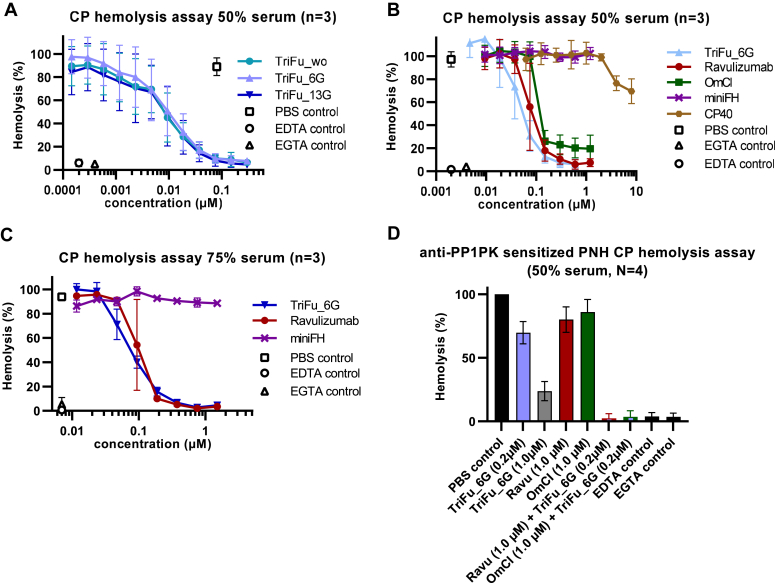
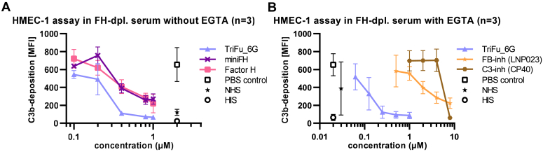
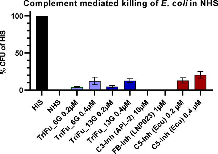
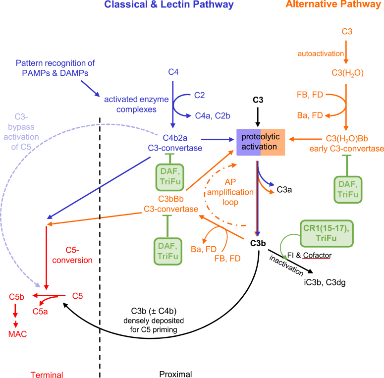
The introduction of a therapeutic anti-C5 antibody into clinical practice in 2007 inspired a surge into the development of complement-targeted therapies. This has led to the recent approval of a C3 inhibitory peptide, an antibody directed against C1s and a full pipeline of several complement inhibitors in preclinical and clinical development. However, no inhibitor is available that efficiently inhibits all three complement initiation pathways and targets host cell surface markers as well as complement opsonins. To overcome this, we engineered a novel fusion protein combining selected domains of the three natural complement regulatory proteins decay accelerating factor, factor H and complement receptor 1. Such a triple fusion complement inhibitor (TriFu) was recombinantly expressed and purified alongside multiple variants and its building blocks. We analyzed these proteins for ligand binding affinity and decay acceleration activity by surface plasmon resonance. Additionally, we tested complement inhibition in several in vitro/ex vivo assays using standard classical and alternative pathway restricted hemolysis assays next to hemolysis assays with paroxysmal nocturnal hemoglobinuria erythrocytes. A novel in vitro model of the alternative pathway disease C3 glomerulopathy was established to evaluate the potential of the inhibitors to stop C3 deposition on endothelial cells. Next to the novel engineered triple fusion variants which inactivate complement convertases in an enzyme-like fashion, stoichiometric complement inhibitors targeting C3, C5, factor B, and factor D were tested as comparators. The triple fusion approach yielded a potent complement inhibitor that efficiently inhibits all three complement initiation pathways while targeting to surface markers.

Keywords: complement; complement receptor 1; compstatin; decay accelerating factor; eculizumab; factor H; fusion protein; paroxysmal nocturnal hemoglobinuria.


Conflict of interest statement

Conflict of interest C. Q. S., H. S., B. H., A. D. and M. H.-L. are inventors of (a) patent application(s) that describes the use of complement inhibitors for therapeutic applications. C. Q. S. has received research funding from pharmaceutical companies. M. H.-L., C. Q. S., B. H. and H. S. received honoraria for speaking at symposia organized by Alexion Pharmaceuticals. H. S. served on advisory committees for Alexion AstraZeneca Rare Diseases, Sanofi, Sobi, Novartis, Amgen and received research funding from Alexion Pharmaceuticals (all to the University of Ulm). The other authors have disclosed no relevant conflict of interest with the contents of the article. B. K. G. and R. B. were/are employees at Takeda Pharmaceuticals.

Figures

Figure 1
Figure 1
C3b and C4b binding affinity of TriFu_6G.A, TriFu_6G was applied onto a carboxymethyl dextran sensor chip surface (CMD500m from Xantec Bioanalytics on a Reichert SR7500DC SPR instrument at 25 °C) with 2070 RU of C3b immobilized via standard amine coupling. The left display shows the sensorgrams of the concentration series. The right display shows the corresponding response versus concentration plot and the 1:1 affinity fit. B, as in (A) but TriFu_6G was assayed for C4b binding with 4820 RUs of C4b being immobilized via amine coupling onto a carboxymethyl dextran sensor chip surface (CMD500m from Xantec Bioanalytics measured on a Reichert SR7500DC SPR instrument at 25 °C). CMD, carboxymethyl dextran; RU, relative unit; SPR, surface plasmon resonance; TriFu, triple fusion complement inhibitor.
Figure 2
Figure 2
Inhibition of AP-mediated hemolysis by TriFu variants and comparison to stoichiometric complement inhibitors.A, TriFu versions with three different linker length were tested in a standard AP-mediated hemolysis assay using rabbit erythrocytes in a final serum content of 50% serum. Lysis in H2O served as a control for 100% lysis and was used to normalize the data. Mean values of three independent assays with standard deviation are shown. B, Trifu_6G was compared to miniFH and eculizumab. Same assay as in (A). Mean values of three independent assays with standard deviation are shown. C, standard AP-mediated hemolysis assay in a higher serum content of 50% comparing the activity of TriFu_6G to ravulizumab OmCI and CP40. Mean values of three independent assays with standard deviation are shown. D, PNH hemolysis assay with a final serum content of 75%. Serum (from different donors of unknown blood group) was slightly acidified (as described in methods according to the modified Ham’s test) and mixed with Mg-EGTA prior to supplementation with analytes and PNH erythrocytes. Level of lysis was normalized to lysis that was observed when PBS instead of a complement inhibitor had been added to the acidified serum. Mean values of three independent assays with standard deviation are shown. AP, alternative pathway; FH, factor H; OmCI, Ornithodoros moubata complement inhibitor; PNH, paroxysmal nocturnal hemoglobinuria; TriFu, triple fusion complement inhibitor.
Figure 3
Figure 3
Inhibition of CP-mediated hemolysis by TriFu variants and comparison to stoichiometric inhibitors.A, three different TriFu version with three different linker length were tested in a CP-mediated hemolysis assay using sensitized sheep erythrocytes in (a higher than normal) final serum content of 50% serum. Lysis in H2O served as a control for 100% lysis and was used to normalize the data. Mean values of three independent assays with standard deviation are shown. B, the inhibitory activity of TriFu_6G toward the CP was compared to ravulizumab, OmCI, miniFH, and CP40. Same assay as in (A). Mean values of three independent assays with standard deviation are shown. C, CP-mediated hemolysis assay in a higher serum content of 75% was used to compare the activity of TriFu_6G to miniFH, and ravulizumab. Mean values of three independent assays with standard deviation are shown. D, CP initiated PNH hemolysis assay without serum acidification at a final serum content of 50%. The alloantibody PP1Pk was used to further sensitize the already vulnerable PNH erythrocytes from PNH patients prior to exposing them to complement active serum that had been supplemented with analytes or controls as indicated. The level of lysis was normalized to lysis observed when PBS instead of a complement inhibitor had been added. Mean values of four independent assays with standard deviation are shown. Fig. S8 shows representative flow cytometry data analyzing the type of PNH erythrocytes that survived the assay conditions. CP, classical pathway; FH, factor H; OmCI, Ornithodoros moubata complement inhibitor; PNH, paroxysmal nocturnal hemoglobinuria; TriFu, triple fusion complement inhibitor.
Figure 4
Figure 4
Protection of human endothelial cells from C3 opsonization in an in vitro model of C3 glomerulopathy. Endothelial cells (HMEC-1) were incubated with NHS, HIS, or FH-depleted serum that had been supplemented with analytes as indicated. The final serum content in the assay was 40%. After incubation at 37 °C for 1 h, the supernatant was discarded, and the cells were harvested and analyzed for C3b deposition by flow cytometry using a monoclonal anti-C3d antibody. Mean values of the measured median fluorescent intensity (MFI) of three independent assays with SD are shown. A and B, show data with and without supplementation of EGTA in the assay, respectively. Representative histograms of the flow cytometry data are shown in Fig. S12. FH, factor H; HIS, heat-inactivated serum; HMEC, human microvascular endothelial cells; NHS, normal human serum.
Figure 5
Figure 5
Complement mediated killing of Escherichia coli in NHS.E. coli cells containing a selective marker were incubated for 1 h in NHS supplemented with complement inhibitors or PBS. The final serum content in the assay was 50%. Colony forming units (CFU) were evaluated by colony counting after plating the reactions on selective medium. The resultant CFU were normalized to the CFU obtained when heat inactivated serum (HIS) was used instead of NHS. Mean values of three technical replicates with standard deviation are shown. NHS, normal human serum.
Figure 6
Figure 6
Schematic illustration of the complement cascade and targets of TriFu and its building blocks with complement regulatory function. The complement cascade was adapted from Mannes et al. 2021 and Schmidt et al. 2016. TriFu has decay accelerating activity toward all pathways as the natural regulator DAF. TriFu also has cofactor activity toward all pathways targeting C4b and C3b as the natural regulator CR1 (for simplicity only C3b cofactor activity is shown). Decay acceleration and cofactor activity of TriFu combined with the targeting functionality of Factor H (not shown) results in an efficient inhibitor of all three complement activation pathways: TriFu targets the activated complement proteins of the classical/lectin and alternative pathway that assemble the convertases (C4b, C3b, C4b, C3bBb, and C4b2a) while it does not affect the constituently expressed precursors (C2, C3, C4, and Factor B). CR1, complement receptor 1; DAF, decay accelerating factor; TriFu, triple fusion complement inhibitor.

Similar articles

Cited by

References

    1. Hillmen P., Young N.S., Schubert J., Brodsky R.A., Socié G., Muus P., et al. The complement inhibitor eculizumab in paroxysmal nocturnal hemoglobinuria. N. Engl. J. Med. 2006;355:1233–1243. - PubMed
    1. Legendre C.M., Licht C., Muus P., Greenbaum L.A., Babu S., Bedrosian C., et al. Terminal complement inhibitor eculizumab in atypical hemolytic-uremic syndrome. N. Engl. J. Med. 2013;368:2169–2181. - PubMed
    1. Howard J.F., Utsugisawa K., Benatar M., Murai H., Barohn R.J., Illa I., et al. Safety and efficacy of eculizumab in anti-acetylcholine receptor antibody-positive refractory generalised myasthenia gravis (REGAIN): a phase 3, randomised, double-blind, placebo-controlled, multicentre study. Lancet Neurol. 2017;16:976–986. - PubMed
    1. Pittock S.J., Berthele A., Fujihara K., Kim H.J., Levy M., Palace J., et al. Eculizumab in aquaporin-4-positive neuromyelitis optica spectrum disorder. N. Engl. J. Med. 2019;381:614–625. - PubMed
    1. Lee J.W., Sicre de Fontbrune F., Wong Lee Lee L., Pessoa V., Gualandro S., Füreder W., et al. Ravulizumab (ALXN1210) vs eculizumab in adult patients with PNH naive to complement inhibitors: the 301 study. Blood. 2019;133:530–539. - PMC - PubMed

Publication types

MeSH terms

LinkOut - more resources