Review
. 2024 Mar;45(3):211-223.
doi: 10.1016/j.it.2024.01.006. Epub 2024 Feb 23.

Gut-associated lymphoid tissue: a microbiota-driven hub of B cell immunity

Affiliations
Review

Gut-associated lymphoid tissue: a microbiota-driven hub of B cell immunity

Mats Bemark et al. Trends Immunol. 2024 Mar.

Abstract

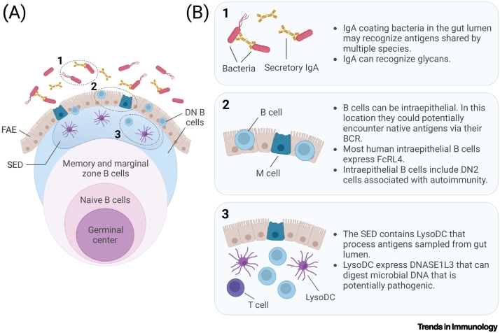
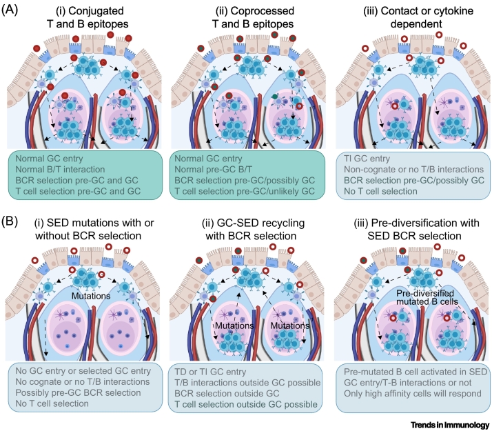
The diverse gut microbiota, which is associated with mucosal health and general wellbeing, maintains gut-associated lymphoid tissues (GALT) in a chronically activated state, including sustainment of germinal centers in a context of high antigenic load. This influences the rules for B cell engagement with antigen and the potential consequences. Recent data have highlighted differences between GALT and other lymphoid tissues. For example, GALT propagates IgA responses against glycans that show signs of having been generated in germinal centers. Other findings suggest that humans are among those species where GALT supports the diversification, propagation, and possibly selection of systemic B cells. Here, we review novel findings that identify GALT as distinctive, and able to support these processes.


Conflict of interest statement

Declaration of interests Michael J. Pitcher and Chiara Dionisi are supported by a Wellcome Trust Investigator award to Jo Spencer (220872/Z/20/Z).

Figures

Figure 1
Figure 1
Example pipeline for processing image data. An example pipeline for processing image data using an imaging mass cytometry example is shown. (A) Raw image files for each region of interest (ROI) are extracted, including cell membrane markers and DNA interlocutor. (B) ROIs are segmented using the image files from (A) to determine cell boundaries within the ROI. (C) Cell measurements are taken including the marker intensity for each cell [from images in (A) and cell boundaries in B)] and cell neighbors based on cell–cell proximities. (D) Cells are assigned identities based on their marker intensities from (C) using, for example, dimensionality reduction and clustering based on key markers. (E) Further analysis is performed, for example visualization of computationally derived cell types back on ROIs and neighborhood analysis finding patches containing similar cell–cell interactions based on assigned phenotypes from (D) and neighbors from (C).
Figure 2
Figure 2
Key figure. Layering of B cells in human gut-associated lymphoid tissue (GALT) with a focus on the subepithelial dome (SED). Schematic representation of human GALT (A) identifying the positions of B cell subsets relative to other key contributors to the follicle-associated epithelium (FAE), which forms the boundary between the microbiota and the host. The germinal center (GC) is a ubiquitous feature of lymphoid tissue in GALT. Surrounding the GC is a zone of naive B cells. This in turn is encircled by the marginal zone and memory B cells. B cells have also been detected in the epithelium [20], where they show expression of Fc receptor-like 4 (FcRL4) [23]. Most B cells adjacent to and within the FAE are double negative (DN) B cells, predominantly of the DN2 subtype, which can accumulate in certain inflammatory and autoimmune diseases [18,25,31,84]. Specialized LysoDCs are present in the SED, where they intermingle with mostly B cells, and handle microbes that are actively sampled by microfold (M) cells in the FAE from the gut lumen [15,17]. LysoDCs contain microbicides and can digest bacterial DNA [16,18]. Above the FAE, secretory IgA-coating bacteria in the gut lumen recognize antigens shared by different species [50], including glycans such as the LPS-O antigen [51]. Areas numbered 1–3 in (A) are illustrated in more detail in (B), along with a description of features that are relevant to this review. This figure was created using Biorender.com.
Figure 3
Figure 3
Potential mechanism supporting T-independent (TI) responses in gut-associated lymphoid tissue (GALT). Gut IgA binding to classical TI antigens carry mutated antibody variable regions, suggesting that responses against TI antigens in GALT differ from those in other tissues. While it is not exactly clear how this is mediated, different mechanisms can be imagined depending on the nature of the antigen. While TI antigens conjugated to proteins act as hapten carriers [33], many gut antigens can be present together, although not covalently linked (i.e., in intact bacteria and viruses). It is therefore possible that T cell epitopes can be presented by B cells that encounter them in the subepithelial dome (SED) [7,21]. Other responses may rely on noncognate interactions with T cells or triggering classical TI responses. Although gut IgA is produced in mice without any germinal centers (GCs) (because of lack of T cell functions), this IgA differs from that in wild-type (WT) mice (lacking variable region mutations); this must be taken into account in any model of TI responses in the gut [34,54]. Abbreviation: Tfh, T follicular helper.
Figure 4
Figure 4
Potential consequences of antigen encounter in gut-associated lymphoid tissue (GALT). (A) For conjugated T-independent (TI) antigens, normal germinal center (GC) responses can occur (i) [33]. If TI and T-dependent (TD) antigens are coprocessed (ii), it is likely that B cells may present epitopes to pre-GC T cells in the subepithelial dome (SED), resulting in cognate interactions and GC entry, but unless intact antigens are transported together to the FDC network, classical GC selection involving T cell interactions cannot occur. For antigens lacking typical T cell epitopes (iii), it is still possible that B cells may enter into already existing GCs because signals required for GC formation and entry into pre-existing GC may differ. (B) Additional processes that might contribute to V region mutations and/or antigen selection in B cells reactive to TI antigens in GALT are mutations in proliferating cells in SED (i) [35,36], B cell receptor (BCR) mediated selection of high-affinity cells leaving GCs to enter the SED during a response (ii) [35,36] or activation of prediversified B cells (i.e., marginal zone B cells in humans or memory B cells with mutations that are not important for binding the original antigen); these may fortuitously be of higher affinity than cells carrying germ-line V regions (iii) [2]. These processes might not be mutually exclusive, but could all contribute.

Similar articles

Cited by

References

    1. Brandtzaeg P., et al. The B-cell system of human mucosae and exocrine glands. Immunol. Rev. 1999;171:45–87. - PMC - PubMed
    1. Weller S., et al. T-independent responses to polysaccharides in humans mobilize marginal zone B cells prediversified against gut bacterial antigens. Sci. Immunol. 2023;8 - PMC - PubMed
    1. Cornes J.S. Number, size, and distribution of Peyer’s patches in the human small intestine: Part I The development of Peyer’s patches. Gut. 1965;6:225–229. - PMC - PubMed
    1. Morbe U.M., et al. Human gut-associated lymphoid tissues (GALT); diversity, structure, and function. Mucosal Immunol. 2021;14:793–802. - PubMed
    1. Perry M., Whyte A. Immunology of the tonsils. Immunol. Today. 1998;19:414–421. - PubMed

LinkOut - more resources