Review
. 2024 Apr;49(4):346-360.
doi: 10.1016/j.tibs.2024.01.011. Epub 2024 Feb 23.

Mitochondria in disease: changes in shapes and dynamics

Affiliations
Review

Mitochondria in disease: changes in shapes and dynamics

Brenita C Jenkins et al. Trends Biochem Sci. 2024 Apr.

Abstract

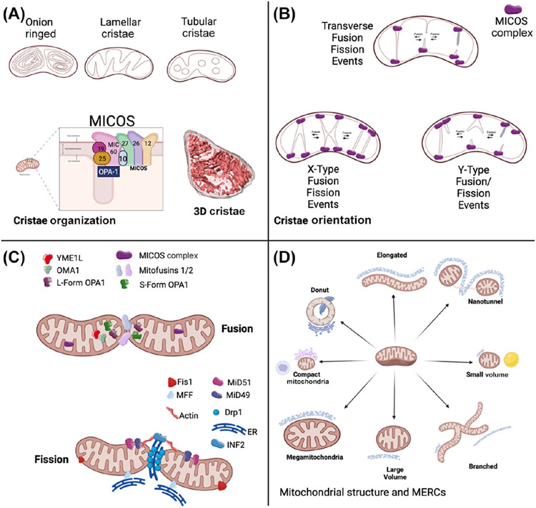
Mitochondrial structure often determines the function of these highly dynamic, multifunctional, eukaryotic organelles, which are essential for maintaining cellular health. The dynamic nature of mitochondria is apparent in descriptions of different mitochondrial shapes [e.g., donuts, megamitochondria (MGs), and nanotunnels] and crista dynamics. This review explores the significance of dynamic alterations in mitochondrial morphology and regulators of mitochondrial and cristae shape. We focus on studies across tissue types and also describe new microscopy techniques for detecting mitochondrial morphologies both in vivo and in vitro that can improve understanding of mitochondrial structure. We highlight the potential therapeutic benefits of regulating mitochondrial morphology and discuss prospective avenues to restore mitochondrial bioenergetics to manage diseases related to mitochondrial dysfunction.

Keywords: clinical diagnostics; contact sites; cristae dynamics; microscopy; mitochondrial morphology; mitochondrial shapes.


Conflict of interest statement

Declaration of interests No interests are declared.

Figures

Figure 3.
Figure 3.
Based on our review, we propose the following essential aspects to consider when researching mitochondrial structures and how their structure may influence their function: cristae types [124]; cristae orientation and fusion/fission events [66,124]; mitochondrial fusion and fission [30,71]; organelle interactions [31]; nucleoid distribution changes [76,77]; tissue-dependent changes [10,11,14,34]; and relative mitochondrial 3D structural phenotypes [15].
Figure 1.
Figure 1.. 3D structures of mitochondria.
3D structures of mitochondria beyond the kidney bean shape, which may have altered cristae dynamics. The eight so-far observed mitochondrial shapes are compact, branching, nanotunnels, mitochondrial donut, elongated, large volume, small volume, and megamitochondria, and these structures, as obtained from 3D reconstructions from serial block face scanning electron microscopy, may have functional implications.
Figure 2.
Figure 2.. Current state of mitochondrial dynamics beyond their phenotype.
(A) Mitochondrial cristae can take on several morphologies, potentially due to the mitochondrial contact site and cristae organizing system (MICOS) complex, and may be imaged in 3D. (B) Mitochondrial cristae can form x- and y-type fusion and fission events, indicating cristae-specific dynamics. (C) Known factors involved in mitochondrial dynamics and potential new regulators. Fission can be mitochondrial fission factor (MFF)- or mitochondrial fission 1 protein (FIS1)-mediated, but the roles of these regulators and others, such as YME1L, in regulating 3D mitochondrial shape remain unclear. (D) Contact sites have several key phenotypes, which may depend on mitochondrial shape. Abbreviations: MERCs, mitochondria–endoplasmic reticulum contact sites; OPA1, optic atrophy 1.

References

    1. Ernster L and Schatz G (1981) Mitochondria: a historical review. J. Cell Biol 91, 227s–255s - PMC - PubMed
    1. Ruska E. (1987) The development of the electron microscope and of electron microscopy. Biosci. Rep 7, 607–629 - PubMed
    1. Palade GE (1952) The fine structure of mitochondria. Anat. Rec 114, 427–451 - PubMed
    1. Palade GE (1953) An electron microscope study of the mitochondrial structure. J. Histochem. Cytochem 1, 188–211 - PubMed
    1. Hackenbrock CR (1966) Ultrastructural bases for metabolically linked mechanical activity in mitochondria. I. Reversible ultrastructural changes with change in metabolic steady state in isolated liver mitochondria. J. Cell Biol 30, 269–297 - PMC - PubMed

Publication types

MeSH terms

LinkOut - more resources