. 2024 Feb 24;19(1):7.
doi: 10.1186/s13008-023-00104-8.

Ultrasonic microbubbles promote mesenchymal stem cell homing to the fibrotic liver via upregulation of CXCR4 expression

Affiliations

Ultrasonic microbubbles promote mesenchymal stem cell homing to the fibrotic liver via upregulation of CXCR4 expression

Heming Xu et al. Cell Div. .

Abstract

Objective: To investigate the mechanism of ultrasound microbubbles (UTMB) promoting stem cells homing to fibrotic liver.

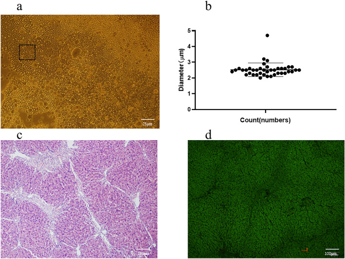
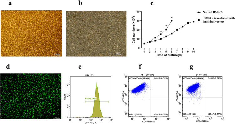
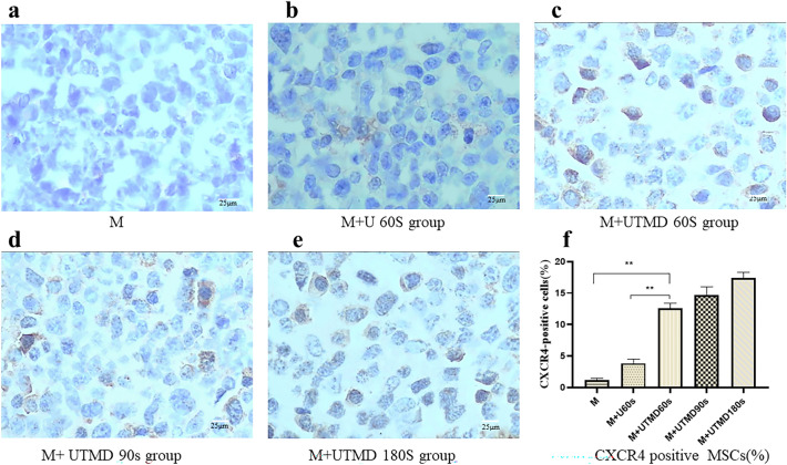
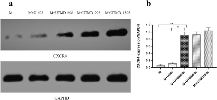
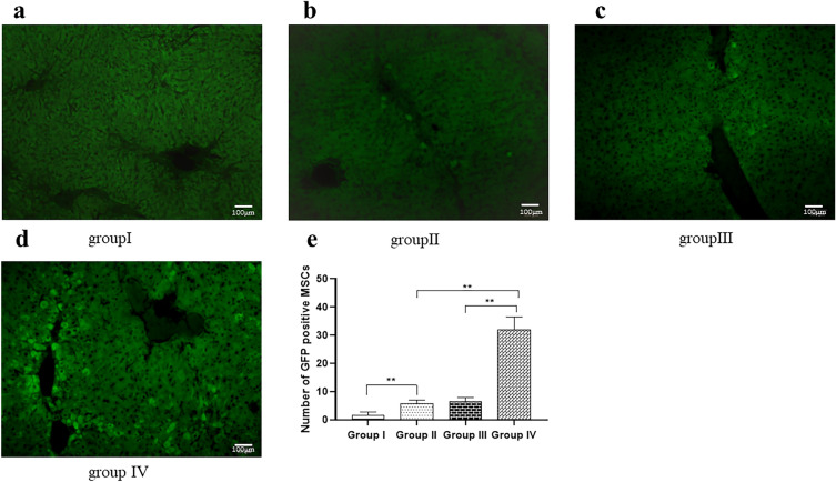
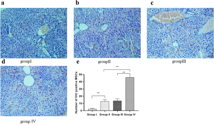
Methods: Bone marrow derived mesenchymal stem cells (BMSCs) were divided into 5 groups with or without ultrasound microbubbles and continuously irradiated with ultrasound conditions of frequency 1 MHZ and output power 0.6 W/cm2 for different times, and then injected into a mouse model of liver fibrosis through the tail vein with or without ultrasound microbubbles, with sound intensity. The effect of ultrasound microbubbles on MSC expression of CXC chemokine receptor 4 (CXCR4) and homing fibrotic liver was evaluated by flow cytometry (FCM), western blot (WB) and immunohistochemistry (IHC) analysis.

Results: The level of CXCR4 expression was significantly higher in the ultrasound microbubble group than in the non-intervention group (P < 0.05), and the number of MSC and the rate of CXCR4 receptor positivity in the ultrasound microbubble-treated liver tissues were significantly higher than in the non-intervention group (P < 0.01).

Conclusion: Ultrasonic microbubbles can promote the expression of CXCR4 on the surface of MSCs, thus improving the homing rate of MSCs in fibrotic liver.

Keywords: CXCR4; Homing; Liver fibrosis; Mesenchymal stem cell; Preface; Ultrasound microbubble.


Conflict of interest statement

The authors declare that the research was conducted in the absence of any commercial or financial relationships that could be construed as a potential conflict of interest.

Figures

Fig. 1
Fig. 1
The identification of microbubble (SonoVue®) and Mouse model. a all microbubbles are less than 5 μm (n = 40, ×100, scale bar = 25 μm). b most microbubble size distribution Between in 2.10–4. 70 μm, the average diameter of microbubbles is about 2.52 ± 0.43 μm. c The mice livers with H&E staining had visible fibrosis, destruction of hepatic lobules, a large amount of hepatocyte apoptosis, and infiltration of inflammatory cells (n = 10, HE ×100, scale bar = 100 μm). d Fluorescence microscopy of HE staining of mouse liver fibrosis
Fig. 2
Fig. 2
Characteristics of mice MSCs (n = 3). a BMSCs transfected with lentiviral vectors were cultured until day 6, The adherent cells reached approximately 80% confluence (×100, scale bar = 100 μm). b Normol BMSCs were cultured until day 6, The adherent cells approximately 100% confluence (×100, scale bar = 100 μm). c The normal BMSCs have better proliferative capacity(*, P < 0.05), Otherwise, The BMSCs transfected with lentiviral vectors can eventually multiply in sufficient numbers. d MSCs were labeled with GFP (×100, scale bar = 100 μm). e FCM revealed that the GFP expression level in BMSCs was approximately 97.21 ± 0.19%. f FCM analysis indicated that the positive rates of MSCs-specific antigens CD29 and CD44 were 97.2 and 97.8%. g The positive rates of CD34 and CD45 were 2.06% and 2.74%
Fig. 3
Fig. 3
Cell surface expression of CXCR4 using FCM (n = 3). ae Representative examples of the membrane expression levels of CXCR4 on MSCs in the different groups. The number of CXCR4-positive cells in the M + UTMB60s group (c) was significantly higher compared with the M group(a) and the M + U60s group (b). There was no significant difference in the number of CXCR4-positive cells between the M + UTMB60s group (c), the M + UTMB90s group (d) and the M + UTMB180s group (e). f Quantification of CXCR4 expression by FCM assay in the experimental MSCs. All values are expressed as the mean ± SD. **P < 0.01; n = 3
Fig. 4
Fig. 4
Expression of CXCR4 by IHC (n = 10). The surface of CXCR4 immunohistochemistry positive cells is brown (× 200, scale bar = 25 μm). ae IHC of CXCR4 in the M group, M + U60s group, M + UTMD 60 s group, M + UTMD 90 s group, M + UTMD 180 s group. The number of CXCR4-positive cells was relatively smaller in the M group (a) and the M + U60s group (b), P > 0.05. The percentage of cells expressing surface CXCR4 in the M + UTMB60s group (c) higher than the M + U60s group (b) and the M group (a), P < 0.05. There was no significant difference in the number of CXCR4-positive cells between the M + UTMB60s group (c), the M + UTMB90s group (d) and the M + UTMB180s group (e), P > 0.05. f Quantification of CXCR4 expression was performed by Image-Pro Plus 6.0 software. All values are expressed as the mean ± SD. **P < 0.01; n = 10
Fig. 5
Fig. 5
Expression of CXCR4 by WB (n = 6). a WB of CXCR4 in the M group, M + U60s group, M + UTMD 60 s group, M + UTMD 90 s group, M + UTMD 180 s group. b Quantification of CXCR4 expression was performed by Image J software. All values are expressed as the mean ± SD. **P < 0.01; n = 6
Fig. 6
Fig. 6
Trypan blue staining for cell viability. The results showed that ultrasound treatment for 60 s and UTMD treatment for 60 s had no significant effect on cell viability. With the further increase of UTMD treatment time from 60 to 180 s, cell viability decreases from 86.33 ± 1.89% to 38.00 ± 1.63% after 48 h of treatment. All values are expressed as the mean ± SD. **P < 0.01; n = 3
Fig. 7
Fig. 7
Representative photograph of GFP staining of MSCs in each group (n = 10). GFP-labeled MSCs were concentrated in the portal vein region under fluorescence microscopy images (×100, scale bar = 100 μm). a There are almost no GFP positive cells in the liver tissue and portal area of the group I. b There are a small number of GFP positive cells in the portal area of the group II. c There are GFP positive cells in the liver tissue and portal area of the group III. d There are a large number of GFP positive cells in the liver tissue and portal area of the group IV. e Quantitative analysis revealed that transplanted MSC in the group II (5.7 ± 1.26) was significantly increased compared to the group I (1.7 ± 1.09) (P < 0.01) and transplanted MSC in the group IV (31.9 ± 4.58) was significantly increased compared with other groups (P < 0.01). All values are expressed as the mean ± SD. **P < 0.01; n = 10
Fig. 8
Fig. 8
Representative photograph of IHC staining of MSCs in each group (n = 10). Anti GFP antibody (ab183734) -labeled MSCs were concentrated in the portal vein region under microscopy images (× 100, scale bar = 100 μm). a a few of IHC positive cells (brown) in the liver tissue and portal area of the group I. b There are a small number of IHC positive cells in the portal area of the group II. c There are IHC positive cells in the liver tissue and portal area of the group III. d There are a large number of IHC positive cells in the liver tissue and portal area of the group IV. e Quantitative analysis revealed that transplanted MSC in the group II (9.8 ± 2.53) was significantly increased compared to the group I (2.4 ± 0.84) (P < 0.01) and transplanted MSC in the group IV (37.1 ± 5.11) was significantly increased compared with other groups (P < 0.01). All values are expressed as the mean ± SD. **P < 0.01; n = 10
Fig. 9
Fig. 9
Expression of SDF-1 by WB (n = 3). a WB of SDF-1 in the Control group, US 60 s group, US + microbubble 60 s group, b Quantification of SDF-1 expression was performed by Image J software. All values are expressed as the mean ± SD. **P < 0.01; n = 3

References

    1. Zhang Y, Li Y, Zhang L, et al. Mesenchymal stem cells: potential application for the treatment of hepatic cirrhosis. Stem Cell Res Ther. 2018;9(1):59. doi: 10.1186/s13287-018-0814-4. - DOI - PMC - PubMed
    1. Terai S, Tsuchiya A. Status of and candidates for cell therapy in liver cirrhosis: overcoming the "point of no return" in advanced liver cirrhosis. J Gastroenterol. 2017;52(2):129–140. doi: 10.1007/s00535-016-1258-1. - DOI - PubMed
    1. Rong X, Liu J, Yao X, et al. Human bone marrow mesenchymal stem cells-derived exosomes alleviate liver fibrosis through the Wnt/β-catenin pathway. Stem Cell Res Ther. 2019;10(1):98–106. doi: 10.1186/s13287-019-1204-2. - DOI - PMC - PubMed
    1. Fang XQ, Liu LW, Dong J, et al. A study about immunomodulatory effect and efficacy and prognosis of human umbilical cord mesenchymal stem cells in patients with chronic hepatitis B-induced decompensated liver cirrhosis. J Gastroenterol Hepatol. 2018;33(4):774–780. doi: 10.1111/jgh.14081. - DOI - PubMed
    1. Hu C, Zhao L, Zhang L, et al. Mesenchymal stem cell-based cell-free strategies: safe and effective treatments for liver injury. Stem Cell Res Ther. 2020;11(1):377–403. doi: 10.1186/s13287-020-01895-1. - DOI - PMC - PubMed

LinkOut - more resources