

A transmission electron microscopy investigation suggests that telocytes, skeletal muscles, myoblasts, and stem cells in common carp (Cyprinus carpio) respond to salinity challenges
- PMID: 38402164
- PMCID: PMC10893627
- DOI: 10.1186/s12917-024-03916-0
A transmission electron microscopy investigation suggests that telocytes, skeletal muscles, myoblasts, and stem cells in common carp (Cyprinus carpio) respond to salinity challenges
Abstract
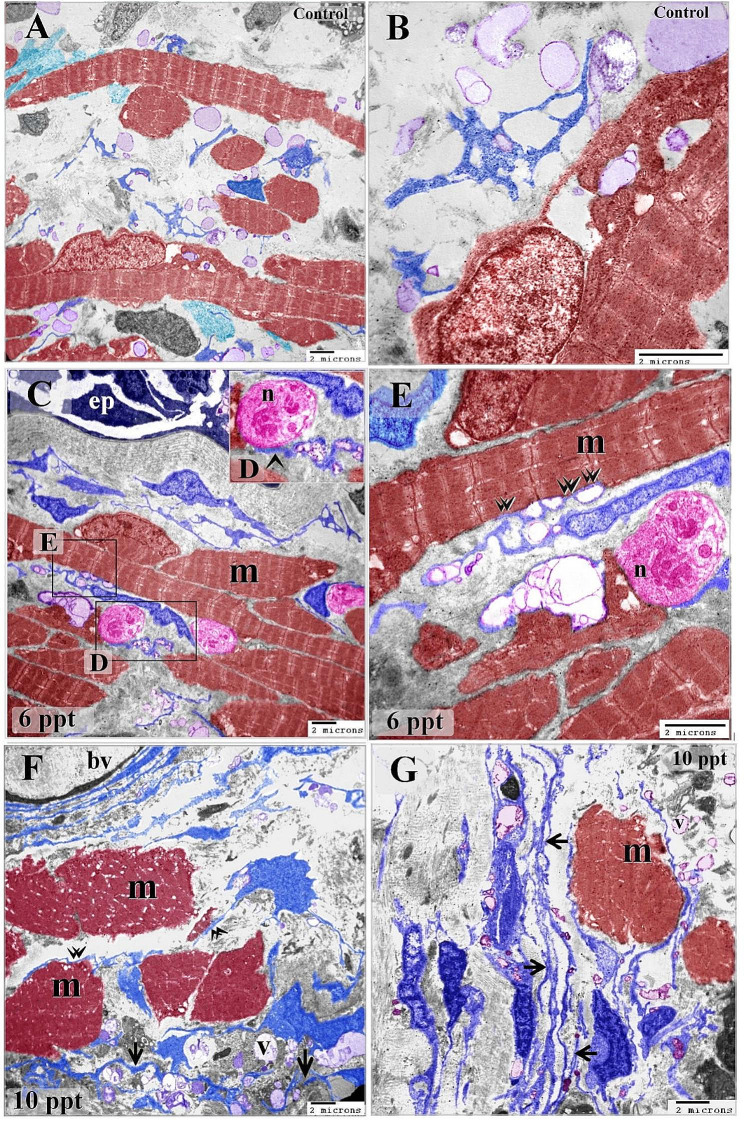
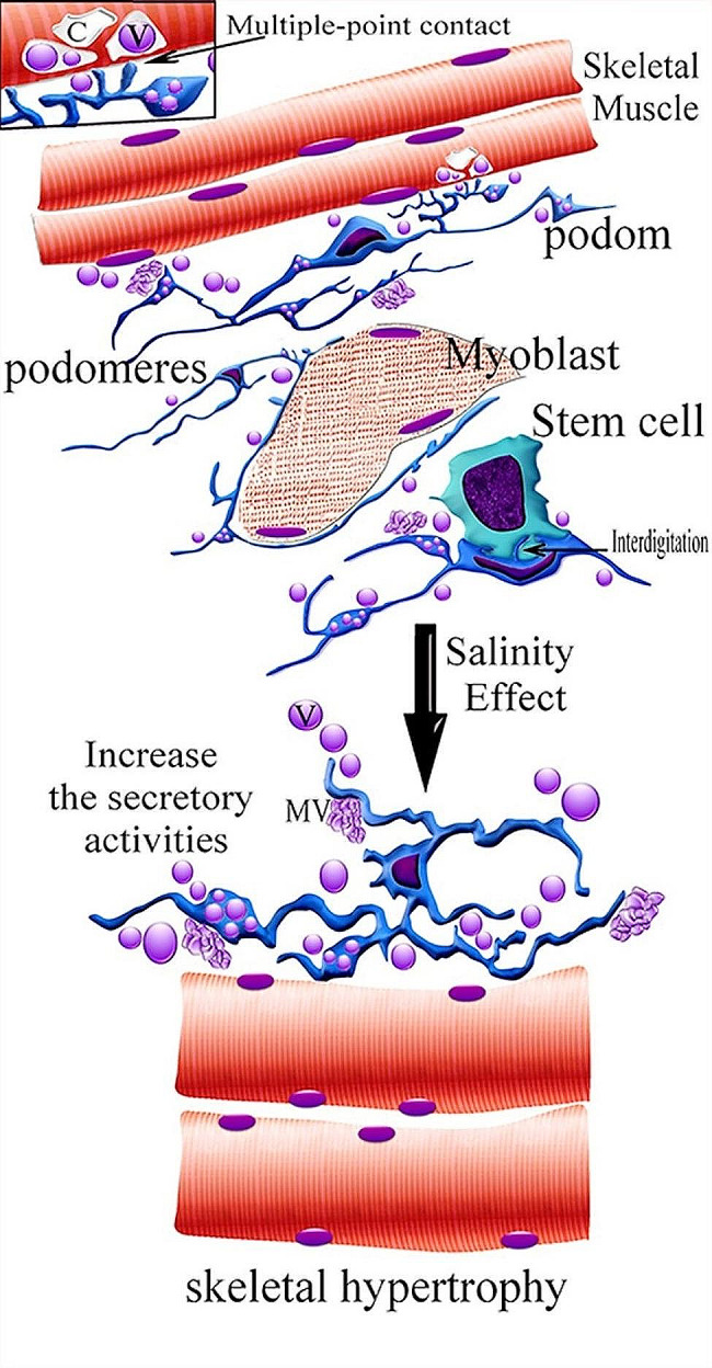
Background: Telocytes are modified interstitial cells that communicate with other types of cells, including stem cells. Stemness properties render them more susceptible to environmental conditions. The current morphological investigation examined the reactions of telocytes to salt stress in relation to stem cells and myoblasts. The common carp are subjected to salinity levels of 0.2, 6, and 10 ppt. The gill samples were preserved and prepared for TEM.
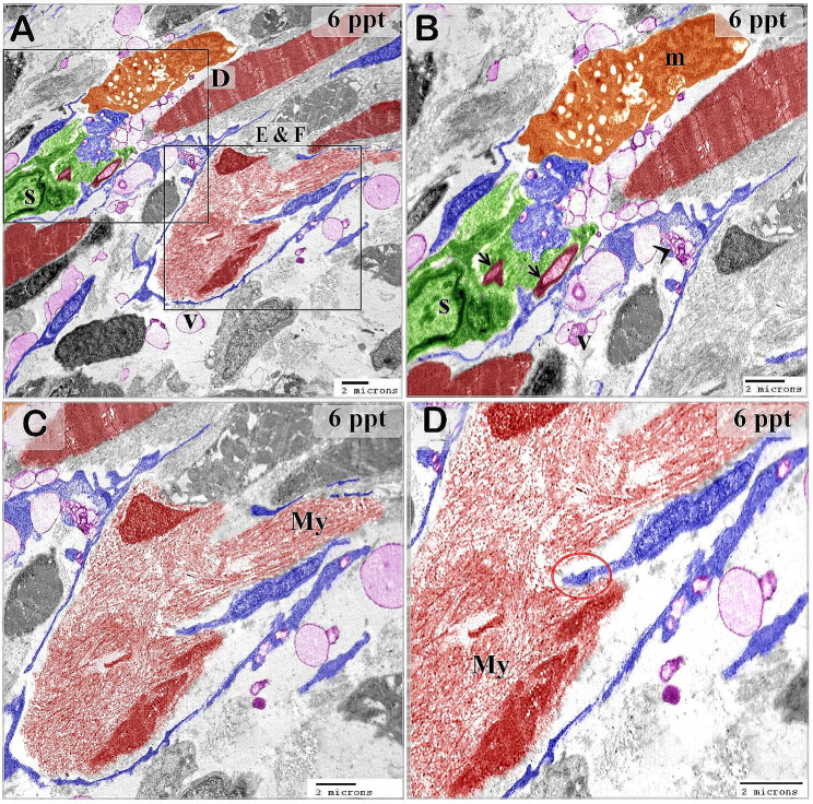
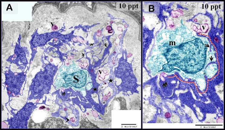
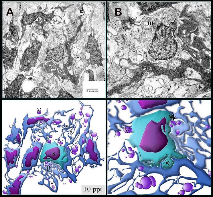
Results: The present study observed that telocytes undergo morphological change and exhibit enhanced secretory activities in response to changes in salinity. TEM can identify typical telocytes. This research gives evidence for the communication of telocytes with stem cells, myoblasts, and skeletal muscles. Telocytes surround stem cells. Telopodes made planar contact with the cell membrane of the stem cell. Telocytes and their telopodes surrounded the skeletal myoblast. These findings show that telocytes may act as nurse cells for skeletal stem cells and myoblasts, which undergo fibrillogenesis. Not only telocytes undergo morphological alternations, but also skeletal muscles become hypertrophied, which receive telocyte secretory vesicles in intercellular compartments.
Conclusion: In conclusion, the activation of telocytes is what causes stress adaptation. They might act as important players in intercellular communication between cells. It is also possible that reciprocal interaction occurs between telocytes and other cells to adapt to changing environmental conditions.
Keywords: Cyprinus carpio; Myoblasts; Salinity; Skeletal muscles; Stem cells; Telocytes.
© 2024. The Author(s).
Conflict of interest statement
The authors declare no competing interests.
Figures


Similar articles
-
Morphological changes in intraepithelial and stromal telocytes in Cyprinus carpio in response to salinity stress.Sci Rep. 2023 Nov 15;13(1):19987. doi: 10.1038/s41598-023-43279-4. Sci Rep. 2023. PMID: 37968439 Free PMC article.
-
A Tale of Two Cells: Telocyte and Stem Cell Unique Relationship.Adv Exp Med Biol. 2016;913:359-376. doi: 10.1007/978-981-10-1061-3_23. Adv Exp Med Biol. 2016. PMID: 27796899 Review.
-
Reappraising the microscopic anatomy of human testis: identification of telocyte networks in the peritubular and intertubular stromal space.Sci Rep. 2018 Oct 3;8(1):14780. doi: 10.1038/s41598-018-33126-2. Sci Rep. 2018. PMID: 30283023 Free PMC article.
-
Identification of telocytes in the porcine heart.Anat Histol Embryol. 2017 Dec;46(6):519-527. doi: 10.1111/ahe.12296. Epub 2017 Sep 7. Anat Histol Embryol. 2017. PMID: 28884484
-
Telocytes: An Emerging Component of Stem Cell Niche Microenvironment.J Histochem Cytochem. 2021 Dec;69(12):795-818. doi: 10.1369/00221554211025489. Epub 2021 Jun 24. J Histochem Cytochem. 2021. PMID: 34165348 Free PMC article. Review.
Cited by
-
Immunohistochemical-properties of the dermal embryonic telocytes.Sci Rep. 2024 Jun 17;14(1):13899. doi: 10.1038/s41598-024-63802-5. Sci Rep. 2024. PMID: 38886354 Free PMC article.
References
-
- Varga I et al. The functional morphology and role of cardiac telocytes in myocardium regeneration. Can J Physiol Pharmacol, 2016: p. 1–5. - PubMed
-
- Rusu MC, et al. Esophageal telocytes and hybrid morphologies. Cell Biol Int. 2012;36(12):1079–88. - PubMed
-
- Mirancea N. Telocyte - a particular cell phenotype. Infrastructure, relationships and putative functions. Rom J Morphol Embryol. 2016;57(1):7–21. - PubMed
MeSH terms
Grants and funding
- grant no. DSR2022-RG-0116./The authors extend their appreciation to the Deanship of Scientific Research at Jouf University for funding this work through research
- grant no. DSR2022-RG-0116./The authors extend their appreciation to the Deanship of Scientific Research at Jouf University for funding this work through research
- grant no. DSR2022-RG-0116./The authors extend their appreciation to the Deanship of Scientific Research at Jouf University for funding this work through research
- grant no. DSR2022-RG-0116./The authors extend their appreciation to the Deanship of Scientific Research at Jouf University for funding this work through research
- grant no. DSR2022-RG-0116./The authors extend their appreciation to the Deanship of Scientific Research at Jouf University for funding this work through research
- grant no. DSR2022-RG-0116./The authors extend their appreciation to the Deanship of Scientific Research at Jouf University for funding this work through research
- grant no. DSR2022-RG-0116./The authors extend their appreciation to the Deanship of Scientific Research at Jouf University for funding this work through research
- grant no. DSR2022-RG-0116./The authors extend their appreciation to the Deanship of Scientific Research at Jouf University for funding this work through research
LinkOut - more resources
Full Text Sources
Research Materials
