. 2024 Feb 24;15(2):171.
doi: 10.1038/s41419-024-06552-6.

DNA polymerase iota promotes EMT and metastasis of esophageal squamous cell carcinoma by interacting with USP7 to stabilize HIF-1α

Affiliations

DNA polymerase iota promotes EMT and metastasis of esophageal squamous cell carcinoma by interacting with USP7 to stabilize HIF-1α

Aidi Gao et al. Cell Death Dis. .

Abstract

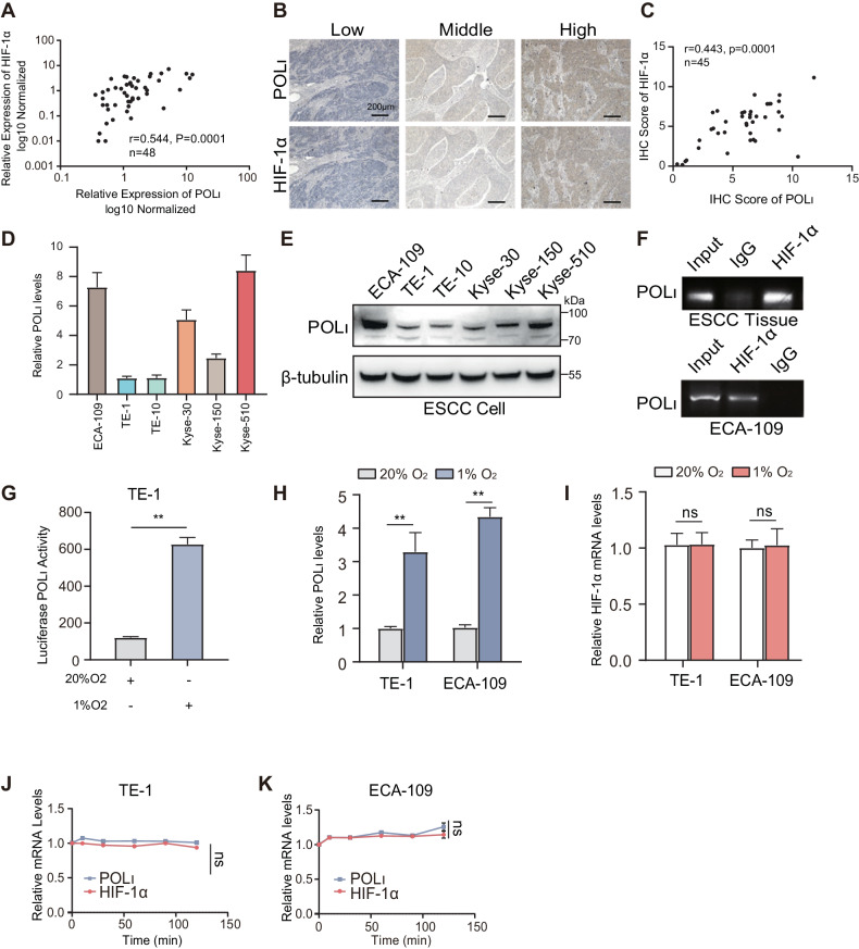
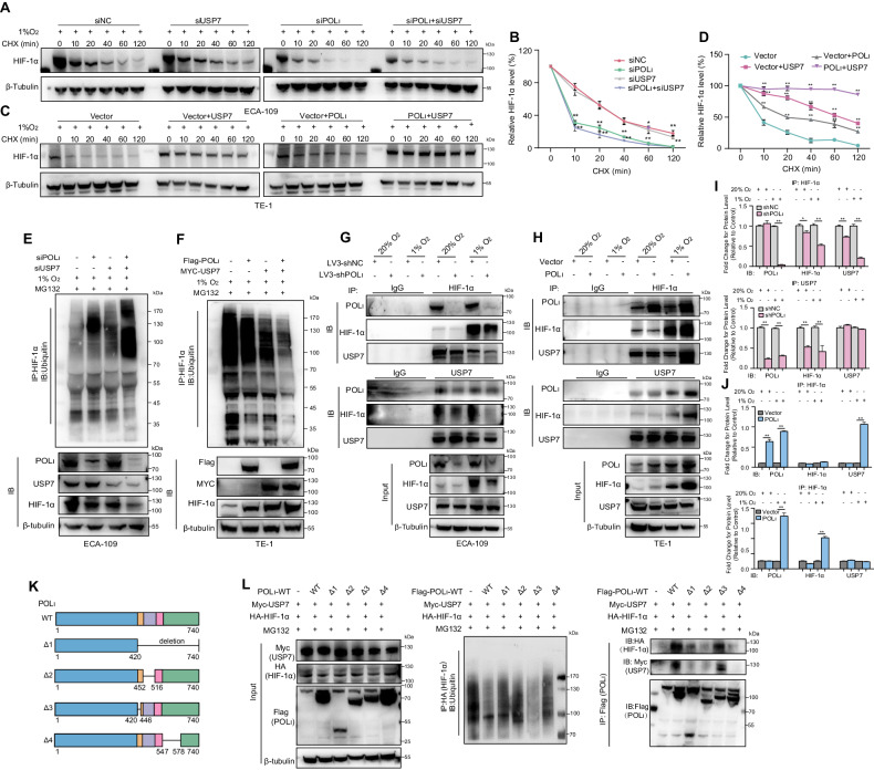
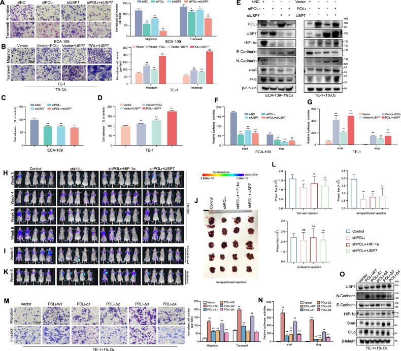
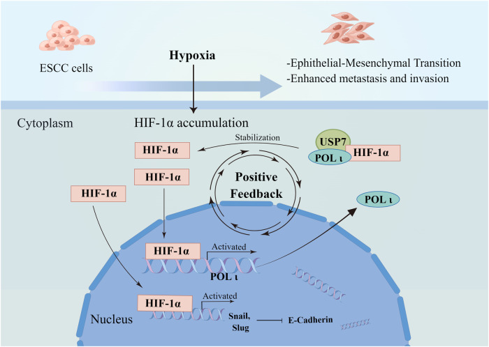
Esophageal squamous cell carcinoma (ESCC) is one of the most lethal cancer types, with a low 5-year survival rate of ~20%. Our prior research has suggested that DNA Polymerase iota (Pol ι), a member of Y-family DNA polymerase, plays a crucial role in the invasion and metastasis of ESCC. However, the underlying mechanism is not well understood. In this study, we utilized ChIP-PCR and luciferase reporter assays to investigate the binding of HIF-1α to the promoter of the Pol ι gene. Transwell, wound healing, and mouse models were employed to assess the impact of Pol ι and HIF-1α on the motility of ESCC cells. Co-immunoprecipitation and Western blot were carried out to explore the interaction between Pol ι and HIF-1α, while qRT-PCR and Western blot were conducted to confirm the regulation of Pol ι and HIF-1α on their downstream targets. Our results demonstrate that HIF-1α activates the transcription of the Pol ι gene in ESCC cells under hypoxic conditions. Furthermore, the knockdown of Pol ι impeded HIF-1α-induced invasion and metastasis. Additionally, we found that Pol ι regulates the expression of genes involved in epithelial-mesenchymal transition (EMT) and initiates EMT through the stabilization of HIF-1α. Mechanistically, Pol ι maintains the protein stability of HIF-1α by recruiting USP7 to mediate the deubiquitination of HIF-1α, with the residues 446-578 of Pol being crucial for the interaction between Pol ι and USP7. Collectively, our findings unveil a novel feedforward molecular axis of HIF-1α- Pol ι -USP7 in ESCC that contributes to ESCC metastasis. Hence, our results present an attractive target for intervention in ESCC.


Conflict of interest statement

The authors declare no competing interests.

Figures

Fig. 1
Fig. 1. HIF-1α activated the transcription of Pol ι in ESCC.
A Correlation analysis of POL ι and HIF-1α mRNA expression in Human ESCC tissue (48 cases). B Immunohistochemistry staining for POL ι and HIF-1α in ESCC specimens (45 cases, 100×). C Correlation analysis of POL ι and HIF-1α protein expression in ESCC tissue with Pearson’s chi-square test. D qPCR analysis of Pol ι mRNA expression in ESCC cell lines. E Western blot analysis of Pol ι protein expression in ESCC cell lines. F Chromatin immunoprecipitation was conducted to assess the binding of HIF-1α to POL ι promoter region in ESCC tissues and in ECA-109 cells. G Pol ι promoter reporter luciferase activity was measured using the dual-luciferase reporter assay. H qPCR analysis of Pol ι mRNA expression in TE-1 and ECA-109 cells under normoxic and hypoxic conditions. I qPCR analysis of HIF-1α mRNA expression after exposure to 20% O2 or 1%O2. J, K The mRNA stability of Pol ι and HIF-1α in TE-1 and ECA-109 cells was determined under normoxic and hypoxic conditions (n = 3, bar, SD). The data were presented as mean ± SD with statistical significance denoted as *P < 0.05; **P < 0.01; and ns, P ≥ 0.05.
Fig. 2
Fig. 2. Pol ι contributes to HIF-1α induced ESCC invasion and metastasis in vitro and in vivo.
A The levels of POL ι and HIF-1α protein were examined by western blot in ECA-109 cells. B Wound healing assay and transwell assays were performed to evaluate the migration and invasion ability of ECA-109 cells with or without overexpression of HIF-1α or knockdown of POL ι. C, D Quantitative analysis of migration and invasion abilities of ECA-109 cells. E Fluorescence imaging of live, lung and kidney from mice injected with ECA-109 cells via tail vein. F The average number of surface nodules per lung was calculated for each group of mice. G Representative images of HE staining of the lung metastatic nodules (40× and 200×). H Immunohistochemical analysis of POL ι and HIF-1α expression in the lung tissues from the mice. I Western blot analysis of POL ι and HIF-1α protein expression in POL ι overexpressed cells and POL ι knockout cells under hypoxic or normoxic. J, K qPCR analysis of POL ι and HIF-1α expression in POL ι overexpressed cells and knockout cells under hypoxic and normoxic. The data were presented as mean ± SD with statistical significance denoted as *P < 0.05; **P < 0.01; and ns, P ≥ 0.05.
Fig. 3
Fig. 3. Pol ι initiates EMT by elevating HIF-1α and its downstream genes under hypoxic in ESCC cells.
A Giemsa staining of ECA-109-sgNC/ECA-109-sgPOLι cells and TE-1-NC/TE-1-POLι cells that were cultured under 20% O2 or 1% O2 conditions for 12 h (magnification: 400×). B Immunofluorescence staining for E-Cadherin, POL ι, and HIF-1α expression in ESCC cells under normoxic and hypoxic. C Transwell assay was performed to evaluate the cell migration and invasion ability. D, E The statistical results of transwell assays. F The adhesive ability of the ESCC cells was assessed by an adhesion assay. The data were presented as mean ± SD with statistical significance denoted as *P < 0.05; **P < 0.01; and ns, P ≥ 0.05. G, H EMT-related protein markers were analyzed using Western blot in ECA-109 cells and TE-1 cells after exposure to normoxic or hypoxic for 12 h.
Fig. 4
Fig. 4. Pol ι inhibits the degradation of HIF-1α through the USP7-mediated deubiquitylation.
A CHX treatment and western blot assay were conducted to measure HIF-1α protein stability in ECA-109 cells. Cells were incubated with 1% O2 for 12 h and then incubated in the presence of CHX for 0, 10, 30, 60,120, or 180 min. B Protein half-life curves were made based on the data. Data were presented as mean ± SD (n = 3). C A CHX chase assay was performed to analyze the half-life of the HIF-1α protein in TE-1 cells. D Protein half-life curves were made based on the data. Data were presented as mean ± SD (n = 3). E, F HIF-1α ubiquitination was determined by Western blot with anti-ubiquitin antibody in ECA-109 cells after treatment with 1% O2 for 12 h. G HIF-1α ubiquitination was determined by western blot with anti-ubiquitin antibody in TE-1 cells after treatment with 1% O2 for 12 h. H Silver staining was conducted to exhibit POL ι interacted proteins. I Mass spectrum analysis of peptides from POL ι and USP7 protein. J IP-western blot was conducted to determine the interaction between POL ι, USP7, and HIF-1α in HEK293T cells transfected with Flag- POL ι, Myc-USP7, and HA-HIF-1α. K Co-immunoprecipitation of endogenous HIF-1α, POL ι, and USP7 in TE-1 and ECA-109 cells. L, M GST pulldown assay was performed to determine the interaction between POL ι, USP7, and HIF-1α. N Co-immunoprecipitation (co-IP) of endogenous HIF-1α, POL ι, and USP7 in 20% O2 or 1% O2-exposeded TE-1-NC/TE-1-POL ι and ECA-109-shNC/ECA-109-shPOL ι cells.
Fig. 5
Fig. 5. Pol ι enhances the binding of USP7 to HIF-1α for its deubiquitination via residues 446–578aa within the C-terminal of Pol ι.
A CHX and western blot assays were used to measure the HIF-1α protein stability in POL ι and USP7 knockdown ECA-109 cells. B Protein half-life curves were made based on the data in ECA-109 cells. C CHX and western blot assays were used to measure HIF-1α protein stability in POL ι and USP7 overexpressed TE-1 cells. D Protein half-life curves were made based on the data in TE-1 cells. Data were presented as mean ± SD (n = 3). E Western blot analysis of the HIF-1α-immunoprecipitated lysates with the anti-ubiquitin antibody in ECA-109 cells. F Western blot analysis of the HIF-1α-immunoprecipitated lysates with the anti-ubiquitin antibody in TE-1 cells. G, H Co-IP assay was performed to determine the binding ability of USP7, POL ι, and HIF-1α in POL ι/USP7 knockdown or overexpression ESCC cells incubated under 1 or 20% oxygen. I, J Quantitative analysis of the co-IP interaction data. K Construction of POL ι wild-type (WT) and deletion mutants with Flag-tag. L In vivo ubiquitination assay and Co-IP were conducted to detect HIF-1α ubiquitination and the binding of POLι mutants to USP7 in HEK293T cells.
Fig. 6
Fig. 6. Blocking the specific binding of Pol ι and USP7 inhibits HIF-1α induced EMT in ESCC cells.
A Images and statistics of the migrated and invaded ECA-109 cells with Pol ι and USP7 knockdown (left panel). The numbers of normalized cell numbers were quantified (right panel). The data (mean ± SD, n = 3) are expressed as normalized cell numbers. **P < 0.01. B Transwell assay was conducted to evaluate the effects of Pol ι and USP7 co-expression on the migration and invasion of TE-1 cells. The numbers of normalized cell numbers were quantified (right panel). Representative images and statistical analysis. The data (mean ± S.D, n = 3) are expressed as normalized cell numbers. **P < 0.01. C, D Adhesion assays to examine adhesion capacity of Pol ι and USP7 knockdown/overexpression ECA-109 and TE-1 cells. E The EMT-related protein levels of Pol ι, USP7, HIF-1α, and EMT-related markers in ECA-109 and TE-1 cells were detected by western blot. F, G Promoter activities of the Snail and Slug genes in ECA-109 and TE-1 cells was measured using a Dual-Luciferase Reporter Assay. The data are presented as mean ± SD with statistical significance denoted as *P < 0.05; **P < 0.01; and ns, P ≥ 0.05. H Live animal imaging of mice at various time points after tail vein injection of ECA-109 cells expressing GFP. I In vivo imaging of mice after intraperitoneal inoculation of ECA-109 cells expressing GFP. J Mice showed liver and spleen metastases after intraperitoneal injection with ECA-109 cells expressing luciferase. K In vivo imaging of mice injected with ECA-109 cells expressing GFP via the left armpit. L Results of quantitative and statistical analyses of the bioluminescence signal intensities in the NC, shPOLι, shPOLι + HIF-1α, and shPOLι + USP7 groups. M Representative image of the transwell assay to evaluate the migration and invasion ability of TE-1 cells expressing different POLι mutants (right panel). The quantitative and statistical analyses are based on normalized cells per field (left panel). N Luciferase reporter assays were used to measure the snail and slug promoter activities in TE-1 cells transfected with empty vector, POLι-WT, and POLι-Δ1–4 plasmids. (n = 3, bar, SD). O EMT-associated protein expression in POLι mutants was analyzed through a western blot assay.
Fig. 7
Fig. 7. Diagram of the feedforward loop of the HIF-1α-POLι-USP7 regulatory axis.
The figure was created using Figdraw.

References

    1. Sung H, Ferlay J, Siegel RL, Laversanne M, Soerjomataram I, Jemal A, et al. Global cancer statistics 2020: GLOBOCAN estimates of incidence and mortality worldwide for 36 cancers in 185 countries. CA Cancer J Clin. 2021;71:209–49. doi: 10.3322/caac.21660. - DOI - PubMed
    1. Siegel RL, Miller KD, Jemal A. Cancer statistics, 2019. CA Cancer J Clin. 2019;69:7–34. doi: 10.3322/caac.21551. - DOI - PubMed
    1. Baiu I, Backhus L. Esophageal cancer surgery. JAMA. 2020;324:1580. doi: 10.1001/jama.2020.2101. - DOI - PubMed
    1. Xia C, Dong X, Li H, Cao M, Sun D, He S, et al. Cancer statistics in China and United States, 2022: profiles, trends, and determinants. Chin Med J. 2022;135:584–90. doi: 10.1097/CM9.0000000000002108. - DOI - PMC - PubMed
    1. Zhou J, Zhang S, Xie L, Liu P, Xie F, Wu J, et al. Overexpression of DNA polymerase iota (Poliota) in esophageal squamous cell carcinoma. Cancer Sci. 2012;103:1574–9. doi: 10.1111/j.1349-7006.2012.02309.x. - DOI - PMC - PubMed

Publication types

MeSH terms