. 2024 Feb 24;15(1):1696.
doi: 10.1038/s41467-024-45806-x.

Prior flavivirus immunity skews the yellow fever vaccine response to cross-reactive antibodies with potential to enhance dengue virus infection

Affiliations

Prior flavivirus immunity skews the yellow fever vaccine response to cross-reactive antibodies with potential to enhance dengue virus infection

Antonio Santos-Peral et al. Nat Commun. .

Abstract

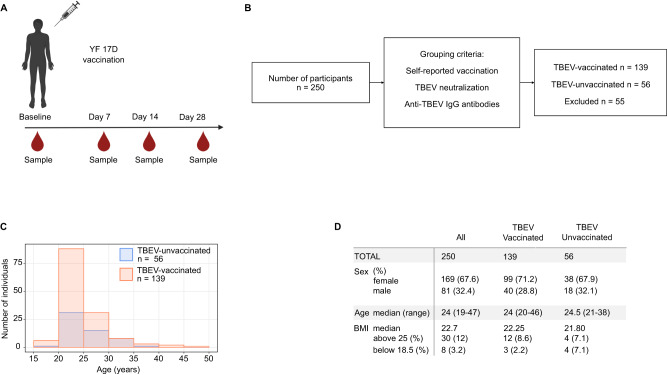
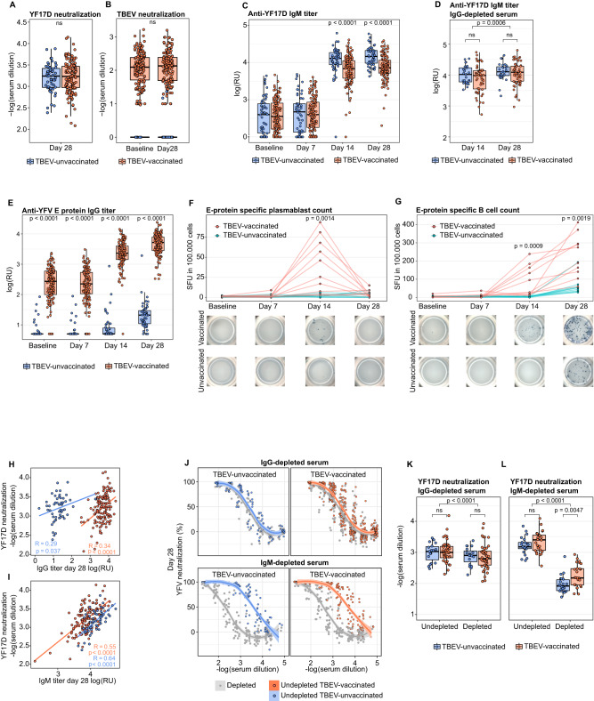
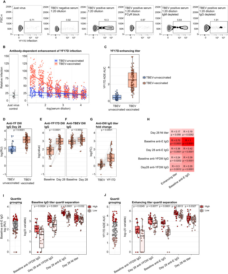
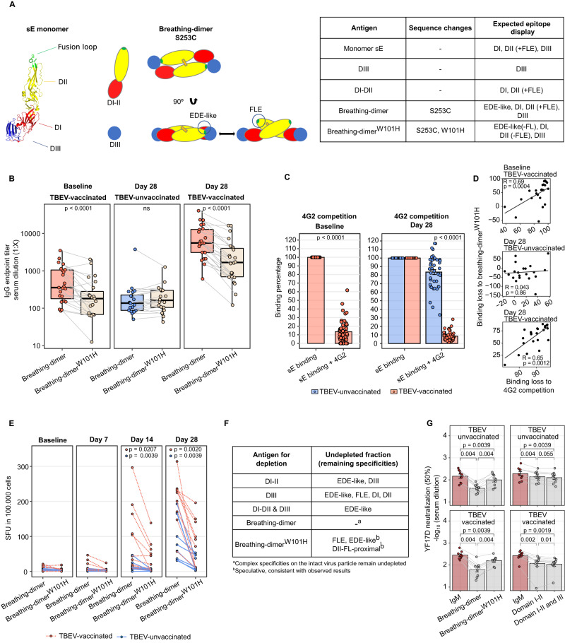
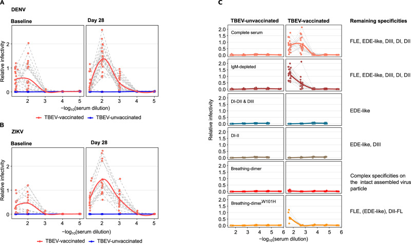
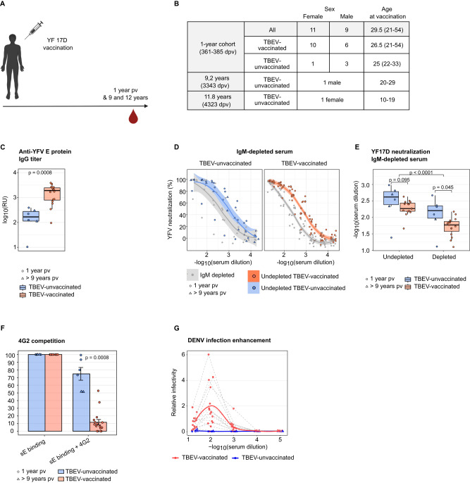
The yellow fever 17D vaccine (YF17D) is highly effective but is frequently administered to individuals with pre-existing cross-reactive immunity, potentially impacting their immune responses. Here, we investigate the impact of pre-existing flavivirus immunity induced by the tick-borne encephalitis virus (TBEV) vaccine on the response to YF17D vaccination in 250 individuals up to 28 days post-vaccination (pv) and 22 individuals sampled one-year pv. Our findings indicate that previous TBEV vaccination does not affect the early IgM-driven neutralizing response to YF17D. However, pre-vaccination sera enhance YF17D virus infection in vitro via antibody-dependent enhancement (ADE). Following YF17D vaccination, TBEV-pre-vaccinated individuals develop high amounts of cross-reactive IgG antibodies with poor neutralizing capacity. In contrast, TBEV-unvaccinated individuals elicit a non-cross-reacting neutralizing response. Using YF17D envelope protein mutants displaying different epitopes, we identify quaternary dimeric epitopes as the primary target of neutralizing antibodies. Additionally, TBEV-pre-vaccination skews the IgG response towards the pan-flavivirus fusion loop epitope (FLE), capable of mediating ADE of dengue and Zika virus infections in vitro. Together, we propose that YF17D vaccination conceals the FLE in individuals without prior flavivirus exposure but favors a cross-reactive IgG response in TBEV-pre-vaccinated recipients directed to the FLE with potential to enhance dengue virus infection.


Conflict of interest statement

The authors declare no competing interests. This work was supported by FlavImmunity a combined grant of the German Research foundation (DFG) project number 391217598 to SR and ABK and the French National Research Agency (ANR) project number ANR-17-CE15-0031-01 to GBS; by DFG TRR237 grant project number 369799452 to SR and ABK (TRR237 TPB14) and BMK (TRR237 TPA04); by Yellow4Flavi a horizon Europe grant project number 101137459 funded by the European Union to SR, AK GBS, KS; by grants of the iMed consortium of the German Helmholtz Societies to SR; by the Einheit für Klinische Pharmakologie (EKLIP), Helmholtz Zentrum München, Neuherberg, Germany to SR and SE; KS and EMS are supported by the BMBF, project 01KI2013; a Stipend (TI 07.003) by the German Center for Infection Research (DZIF) to FL; grants by the Friedrich Baur Foundation (FBS) to JTS, HK and MP; a Metiphys fellowship of the Medical Faculty of the LMU Munich to MP; by the FöFoLe Program of the Medical Faculty of the LMU Munich to SG, EN, LL, FL, the international doctoral program “iTarget: Immunotargeting of cancer” funded by the Elite Network of Bavaria to ASP, MZ, SG, LL, EN. The funders had no role in study design, data collection and analysis, decision to publish, or preparation of the manuscript.

Figures

Fig. 1
Fig. 1. Cohort-1. Overview of study participants.
A Diagram representing the longitudinal PBMC, serum, and plasma sample collection of 250 participants. Samples were collected at baseline (immediately before vaccination) and on days 7, 14 and 28 post vaccination. Prepared with Biorender (www.biorender.com) B Flow chart of cohort members grouping according to TBEV pre-vaccination status. 139 individuals self-reported having received at least one TBEV-vaccine dose and contained neutralizing antibodies and anti-TBEV IgG at baseline. 56 TBEV-unvaccinated individuals were classified based on a self-reported negative vaccination history validated by the absence of detectable anti-TBEV IgG and neutralizing capacity at baseline C Histogram depicting the age distribution of the 139 TBEV-vaccinated participants (in orange) and the 56 TBEV-unvaccinated donors (in blue). D Table summarizing cohort-1 characteristics for all 250 individuals and separated according to TBEV-pre-vaccination status.
Fig. 2
Fig. 2. Comparison of the YF17D-induced antibody and B cell responses between TBEV-vaccinated and unvaccinated participants.
A YF17D 80% neutralization titer at day 28 post-vaccination for TBEV-vaccinated (n = 137) and unvaccinated (n = 56) individuals. B TBEV 80% neutralization titer before and 28 days after YF17D vaccination for TBEV-vaccinated individuals (n = 137) and TBEV-unvaccinated individuals (n = 56). C Longitudinal YF17D virion-specific IgM response in TBEV pre-vaccinated (baseline, n = 132; day 7, n = 128; day 14, n = 128; day 28, n = 129) and TBEV unvaccinated donors (baseline, n = 54; day 7, n = 54; day 14, n = 54; day 28, n = 53). D YF17D virion-specific IgM titer on days 14 and 28 in IgG-depleted serum for TBEV pre-vaccinated (n = 56) and TBEV unvaccinated donors (n = 28). E Longitudinal YF17D anti-E protein-specific IgG titers for TBEV-vaccinated (baseline, n = 136; day 7, n = 131; day 14, n = 132; day 28, n = 133) and TBEV-unvaccinated donors (baseline, n = 52; day 7, n = 52; day 14, n = 52; day 28, n = 52). F, G Plasmablasts and total sE-specific longitudinal B-cell response quantified by ELISpot and depicted as spot-forming units (SFU) in 100,000 PBMC (n = 10 TBEV-vaccinated and n = 9 TBEV-unvaccinated donors). Spot pictures are shown for a representative example of a TBEV-pre-vaccinated and unvaccinated individual. Significance compares B cell counts between groups on days 14 (F) and 28 (G). H, I Spearman correlation between the YF17D polyclonal neutralizing titer of sera on day 28 with the IgG titer (TBEV-vaccinated n = 133 and TBEV-unvaccinated donors n = 52) and IgM titer (TBEV-vaccinated n = 129, TBEV-unvaccinated donors n = 53). J Neutralization curves of undepleted polyclonal serum and IgG or IgM-depleted serum (in grey) for TBEV-pre-vaccinated (n = 45 and 26, respectively) and unvaccinated individuals (n = 19 and n = 22, respectively). Quantification of the 80% neutralization cutoff before and after IgG (K) and IgM (L) depletions shown in J. TBEV-vaccinated participants are depicted in orange and TBEV-unvaccinated in blue. Boxplots show a horizontal line indicating the median and lower and upper hinges corresponding to the first and third quartiles. The lower and upper whiskers extend to 1.5x IQR (interquartile range) from the respective hinge. The curve fitting in J was done with local regression with a 0.95 confidence interval. Statistical significance between TBEV-vaccinated and unvaccinated individuals is shown above every comparison and was estimated with a two-sided Mann-Whitney test. Statistical significance between undepleted and depleted samples (K and I) and between timepoints (D) was calculated with a two-sided Wilcoxon signed-rank test. P values above 0.05 are considered non-significant (ns).
Fig. 3
Fig. 3. Antibody dependent enhancement of YF17D virus infection mediated by TBEV vaccine-induced IgG.
A Flow cytometric determination of venus-YF17D virus infection of THP-1 cells in the absence or presence of cross-reactive serum from a TBEV-vaccinated individual. The conditions tested include polyclonal serum alone or in combination with FcγR-blocking antibodies and IgM or IgG-depleted serum. B ADE of YF17D mediated by study participants’ serum (n = 132 TBEV-vaccinated, n = 53 TBEV-unvaccinated individuals). Virus infection was quantified in combination with serially diluted serum and was normalized against the enhancement of a 1:20 diluted enhancing-serum control carried for all measurements. Thick dashed line indicates the mean of the different just-virus controls and dotted lines define +/− 1 SD. The curve was fitted with local regression with a 0.95 confidence interval. C Quantification of the enhancing titer as AUC of the normalized virus infection across serial dilutions shown in B. D Comparison of anti-YF17D-DIII specific IgG titer at day 28 post-vaccination for both TBEV groups (n = 36 TBEV-unvaccinated, n = 117 TBEV-vaccinated individuals). Longitudinal quantification of anti-YF17D-DIII (E, n = 97 donors) and anti-TBEV-DIII (F, n = 114 donors) specific IgG in TBEV-experienced individuals. G Paired comparison of the DIII-specific IgG fold-change between day 28 and day 0 for TBEV and YF17D (n = 76 pairs). H Spearman correlation in TBEV-experienced individuals of baseline anti-E IgG and enhancing titers versus YF17D vaccine-induced neutralizing antibody titer at day 28, anti-sE and anti-DIII IgG titers and baseline anti-YF17D-E and anti-YF17D-DIII IgG titers. Color intensity reflects the Spearman correlation coefficient. I Comparison of the first (n = 34) and fourth (n = 33) quartile group of YF17D-sE specific IgG at baseline and the anti-YFDIII IgG on baseline and day 28, the anti-YF17D-sE IgG titer on day 28 and the anti-YF17D neutralizing antibody titer on day 28 (high vs. low quartile n = 24/23, 32/28, 33/32, 34/33 respectively). J Comparison of the first (n = 33) and fourth (n = 33) quartile group of the Enhancing titers at baseline and the anti-YFDIII IgG on baseline and day 28, the anti-YF17D-sE IgG titer at baseline and day 28 and the anti-YF17D neutralizing antibody titer on day 28 (high vs low quartile n = 25/22, 28/30, 32/33, 32/33, 33/33, respectively). TBEV-vaccinated participants are depicted in orange and TBEV-unvaccinated in blue. Boxplots show a horizontal line indicating the median and lower and upper hinges corresponding to the first and third quartiles. The lower and upper whiskers extend to 1.5x IQR from the respective hinge. Statistical significance between TBEV-vaccinated and unvaccinated individuals (D) and between high and low quartiles (I, J) is shown above every comparison and was estimated with a two-sided Mann-Whitney test. Statistical significance in EG was calculated with a two-sided Wilcoxon signed-rank test. P values above 0.05 are considered non-significant (ns).
Fig. 4
Fig. 4. IgG and IgM cross-reactivity signature before and after YF17D vaccination in TBEV-unvaccinated and pre-vaccinated individuals.
IgG (A) and IgM (B) subtype cross-reactivity evaluation before and after YF17D vaccination using an indirect immunofluorescent test for a panel of nine human-pathogenic flaviviruses: DENV 1–4, ZIKV, WNV, JEV, YFV and TBEV. A subgroup of n = 39 individuals was tested (Fig. S3), out of which n = 15 were TBEV-unvaccinated and n = 24 TBEV-experienced. Bars indicate titer mean and dots reflect the antibody amounts as serum dilution end-point titers. TBEV-vaccinated participants are depicted in orange and TBEV-unvaccinated in blue.
Fig. 5
Fig. 5. Binding sites and neutralizing capacity of the YF17D vaccine-induced IgG response in TBEV-unvaccinated and pre-vaccinated individuals.
A Recombinant proteins for the dissection and functional analysis of different antibody specificities. The illustration depicts the envelope protein ectodomains (sE), DI-II, and DIII produced separately as well as recombinantly produced dimeric structures. The table summarizes the epitopes displayed by the protein antigens used. B IgG endpoint titer quantification for breathing-dimer and breathing-dimerW101H specificities by ELISA at baseline (n = 23 TBEV-vaccinated donors) and day 28 (n = 24 TBEV-vaccinated, n = 20 TBEV-unvaccinated individuals). C Antibody binding competition to sE of participants serum with the FL-mab 4G2 at baseline (n = 55 TBEV-vaccinated) and day 28 (n = 55 TBEV-vaccinated and n = 43 TBEV-unvaccinated donors). The percentage of remaining binding is calculated by comparing the binding signal with and without 4G2 as competitor D Spearman correlation between antibody binding loss estimated with the 4G2 competition assay (C) and with the breathing-dimerW101H (B) (n = 23 pairs of baseline samples, n = 23 pairs of TBEV-vaccinated and n = 20 TBEV-unvaccinated of day 28 samples) E Longitudinal quantification of IgG-producing B-cells specific for breathing-dimer and breathing-dimerW101H (n = 10 TBEV-vaccinated and n = 9 TBEV-unvaccinated donors). Units represent spot-forming units per 100.000 PBMC. F Table describing the antigens used for antigen-specific IgG depletions and the expected specificities of the remaining undepleted fraction used for YF17D neutralization assays. G YF17D neutralization titers (50% cutoff) of IgM-depleted and antigen-specific-depleted sera as explained in F (n = 9 TBEV-vaccinated and n = 9 TBEV-unvaccinated participants). TBEV-vaccinated participants are depicted in orange and TBEV-unvaccinated in blue. Envelope structure accession number (PDB: 6IW4) was edited using Pymol. Individual selection is shown in Supplementary Fig. 3 (SF3). Boxplots display a horizontal line indicating the median and lower and upper hinges corresponding to the first and third quartiles. The lower and upper whiskers extend to 1.5x IQR from the respective hinge. Barplots in C and G indicate the mean and the error bars the standard error of the mean. Statistical significance between TBEV-vaccinated and unvaccinated individuals (C) was estimated with a two-sided Mann-Whitney test. Statistical significance in B, E and G was calculated with a two-sided Wilcoxon signed-rank test. P values above 0.05 are considered non-significant (ns).
Fig. 6
Fig. 6. Antibody-dependent enhancement of DENV and ZIKV by bulk and antigen-specific IgG depleted serum in TBEV-unvaccinated and pre-immunized individuals before and after YF17D vaccination.
A Antibody-dependent enhancement of infection with DENV-2 (16681) VRPs at baseline and day 28 post-YF17D vaccination (n = 23 TBEV-vaccinated, n = 15 TBEV-unvaccinated individuals) B Antibody-dependent enhancement of infection with ZIKV (MR-766 African strain) VRPs at baseline and day 28 post-YF17D vaccination (n = 16 TBEV-vaccinated, n = 8 TBEV-unvaccinated individuals). C Dengue ADE for TBEV-vaccinated and unvaccinated individuals driven by: undepleted serum (n = 22 per group), IgM-depleted serum (n = 22 per group), and, as detailed in Fig. 5F, IgM & antigen-specific-IgG-depleted serum (n = 9 per depletion group). Relative infectivity is estimated as the normalized fold-increase of infection to an internal control carried for all the assays. Curves were fitted with local regression. In A and B TBEV-vaccinated participants are depicted in orange and TBEV-unvaccinated in blue.
Fig. 7
Fig. 7. IgG antibody response in cohort-2 sampled one year after YF17D vaccination.
A Diagram representing the serum sample collection of 22 participants. Prepared with Biorender (www.biorender.com). B Table summarizing cohort-2 characteristics and TBEV-pre-vaccination status. The table indicates the age at the time of vaccination. C YF17D anti-E protein-specific IgG titers in relative units (RU). D Neutralization curves of undepleted polyclonal serum and IgG- or IgM-depleted serum (in grey) for TBEV-pre-vaccinated or unvaccinated individuals. E Quantification of the 80% neutralization cutoff before and after IgM depletion. F Antibody binding competition to sE of participants serum with the FL-mAb 4G2. Percentage of remaining binding is calculated by comparing the binding signal with and without 4G2 as competitor. G Antibody-dependent enhancement of infection with DENV-2 VRPs. TBEV-vaccinated participants (n = 16) are depicted in orange and unvaccinated (n = 6) in blue. One-year pv samples are represented with circles and over 9 years pv with triangles. All individuals are included in every assay except for D and E (n = 15 TBEV-vaccinated donors). Curve fitting in D and G was calculated with local regression. Boxplots display a horizontal line indicating the median and lower and upper hinges corresponding to the first and third quartiles. The lower and upper whiskers extend to 1.5x IQR from the respective hinge. Barplots in F indicate the mean and the error bars the standard error of the mean. Statistical significance between TBEV-vaccinated and unvaccinated individuals (C, E and F) was estimated with a two-sided Mann-Whitney test. Statistical significance between undepleted and IgM-depleted serum samples in E was calculated with a two-sided Wilcoxon signed-rank test. P values above 0.05 are considered non-significant (ns).

Similar articles

Cited by

References

    1. Vasilakis N, Cardosa J, Hanley KA, Holmes EC, Weaver SC. Fever from the forest: prospects for the continued emergence of sylvatic dengue virus and its impact on public health. Nat. Rev. Microbiol. 2011;9:532–541. doi: 10.1038/nrmicro2595. - DOI - PMC - PubMed
    1. Kraemer MUG, et al. The global distribution of the arbovirus vectors Aedes aegypti and Ae. albopictus. Jit M, editor. Elife. 2015;4:e08347. doi: 10.7554/eLife.08347. - DOI - PMC - PubMed
    1. Gould EA, Solomon T. Pathogenic flaviviruses. Lancet. 2008;371:500–509. doi: 10.1016/S0140-6736(08)60238-X. - DOI - PubMed
    1. Pierson TC, Diamond MS. The continued threat of emerging flaviviruses. Nat. Microbiol. 2020;5:796–812. doi: 10.1038/s41564-020-0714-0. - DOI - PMC - PubMed
    1. Stiasny, K., Medits, I., Roßbacher, L., Heinz, F. X. Impact of structural dynamics on biological functions of flaviviruses. FEBS J. 290, 1973–1985 (2022). - PMC - PubMed