. 2024 Feb 24;15(1):1709.
doi: 10.1038/s41467-024-45985-7.

A streamlined approach to structure elucidation using in cellulo crystallized recombinant proteins, InCellCryst

Affiliations

A streamlined approach to structure elucidation using in cellulo crystallized recombinant proteins, InCellCryst

Robert Schönherr et al. Nat Commun. .

Abstract

With the advent of serial X-ray crystallography on microfocus beamlines at free-electron laser and synchrotron facilities, the demand for protein microcrystals has significantly risen in recent years. However, by in vitro crystallization extensive efforts are usually required to purify proteins and produce sufficiently homogeneous microcrystals. Here, we present InCellCryst, an advanced pipeline for producing homogeneous microcrystals directly within living insect cells. Our baculovirus-based cloning system enables the production of crystals from completely native proteins as well as the screening of different cellular compartments to maximize chances for protein crystallization. By optimizing cloning procedures, recombinant virus production, crystallization and crystal detection, X-ray diffraction data can be collected 24 days after the start of target gene cloning. Furthermore, improved strategies for serial synchrotron diffraction data collection directly from crystals within living cells abolish the need to purify the recombinant protein or the associated microcrystals.


Conflict of interest statement

The authors declare no competing interests.

Figures
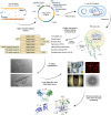
Fig. 1
Fig. 1. The InCellCryst pipeline.
The gene of interest is amplified by PCR and ligated into modified pFastBac1 plasmids. After transformation of E. coli DH10EmBacY cells, recombination with the bacmid takes place. The recombinant bacmid is isolated and Sf9 insect cells are transfected for BV generation. After high titer viral stock production, High Five insect cells are infected and used for high yield target gene expression. This eventually leads to the crystallization of the target protein within one of the cellular compartments, depending on the transport signaling tag fused to the target protein sequence. Crystal-containing cells are directly used for serial diffraction data collection at RT or 100 K at a synchrotron source or an XFEL. Serial diffraction data is finally processed to elucidate the structure of the target protein. rBVs recombinant baculoviruses.
Fig. 2
Fig. 2. Optimization of virus stock production.
a Comparison of the susceptibility of different insect cell lines for recombinant baculovirus (rBV) infection. Three different rBVs were titrated on High Five cells as well as on two Sf9 cell lines obtained from different sources. The averaged TCID50-values of four independent experiments are presented as mean values ± SD. High Five cells exhibit the highest apparent titer due to their increased susceptibility. b Comparison of virus production of High Five and Sf9 cell lines. Two different rBVs (with/ without crystal production capability) were amplified on the denoted cell lines in three independent experiments and titrated twice on High Five cells. For the infection, a titer of 1 × 104 ml−1 was used. The resulting virus stock was harvested 4 days after infection of 0.45 × 106 cells in a 12-well plate. The averaged TCID50-values are presented as mean values±SD. c Comparison of virus productions depending on the initial infection titer. Experiment design as described in b. Infection titers varied between 1 × 101 and 1 × 106 ml−1. The differences between infection and harvesting titers are shown as amplification factors. High Five cells do not produce a noticeable amount of new infectious viral particles, while Sf9 cells are shown to be highly productive. Different clones of the same cell line can exhibit considerable differences in their virus production capabilities. Data of three independent experiments are presented as mean values ± SD.
Fig. 3
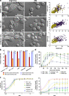
Fig. 3. Crystallization capabilities of insect cell lines.
a Visual comparison of crystals in Sf9 and High Five cells, illustrating the size differences between crystals of the same protein produced in the two cell lines. Representative images of three independent experiments are shown. High Five cells produce significantly larger crystals than Sf9 cells while the morphology remains identical. Measurements of several hundred crystals in the right-side panels illustrate the large size differences. Yellow dots, Sf9 cells; violet dots, High Five cells. b High Five cells also show improved crystallization capabilities compared to Sf9 cells characterized by a higher fraction of crystal-containing cells within an infected culture, while no differences are visible regarding the fraction of infected, EYFP-producing cells. c An MOI of 0.5 is sufficient for infection of and protein production in nearly all cells within a High Five cell culture. However, a MOI of 1 should be used to maximize protein crystallization within the cells. A higher MOI does not improve in cellulo crystallization, independent of the crystallizing protein. d Visible crystal growth in High Five cells is detectable from 36 h after infection with an MOI of 1 onwards. Growth characteristics are variable but follow a comparable scheme with the maximal fraction of crystal-containing cells obtained between 72 and 96 hpi. The only exception is IMPDH cyto, showing a continuous increase in crystal containing cells. bd Data of three independent experiments are presented as mean values ± SD.
Fig. 4
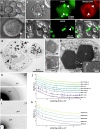
Fig. 4. Methods for detection and analysis of intracellular protein crystals.
a Imaging of HEX-1 SS crystals (white arrow) in High Five cells using differential interference contrast (DIC). b Immunofluorescence labeling of HA-tagged HEX-1 HA-C crystals (white arrows) in High Five cells with a DyLight 549-conjugated antibody. c Confocal fluorescence imaging of EGFP-µNS in Sf9 cells. d TEM of High Five cells producing luciferase+ cyto. Crystals are visible in high contrast due to their comparatively high protein density. e TEM of nanocrystals of a baculoviral protein located within the ER of a Sf9 cell showing a fine crystal lattice grating. f TEM of a CatB crystal surrounded by a ribosome studded membrane (rER) in a Sf9 cell. g TEM of EGFP-µNS crystals in High Five cells showing defects in the crystal lattice. h, i Powder diffraction images of High Five cells containing crystals of HEX-1 cyto h or IMPDH HA-N i. For IMPDH background subtraction was done using adxv to enhance the visibility of Debye-Scherrer rings. j, k SAXS curves of crystal-containing High Five cell suspensions. Peaks arise from incomplete Debye-Scherrer rings on the detector images. Graphs correspond to cells producing HEX-1 j and IMPDH k proteins fused to different tags and localization sequences. In contrast to IMPDH, crystals of HEX-1 variants give diverse fingerprints, implying differences in the unit cell parameters. Cr protein crystal, Cp cytoplasm, ER endoplasmic reticulum, N nucleus, Nc nucleocapsid, rERM membrane of the rough ER. Representative micrographs of three independent experiments are shown.
Fig. 5
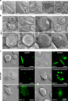
Fig. 5. Crystal morphology depends on the cellular compartment.
rBVs were used to infect High Five a, b or Sf9 cells c at an MOI of 1. Imaging followed 4 dpi on a Nikon Ti2-E or Ts2R-FL microscope equipped with 100x objectives using the DIC contrast mode and EGFP wide-field fluorescence. Size bars for all images represent 20 µm a, b and 15 µm c, respectively. Representative images of three independent experiments are shown. a Compartment screening of IMPDH. Crystallization success and crystal morphology depend on the target organelle. The unmodified IMPDH (ori) as well as variants without the native (cyto) or with an artificial PTS1 motif crystallize within the cytosol, but no crystallization can be observed when the protein is translated into the ER. Crystal morphology is comparable in all compartments that enable crystallization. b Compartment screening of HEX-1. The unmodified HEX-1 crystallizes in blocky hexagons. The N- and C-terminal addition of single amino acids (cyto) leads to spindle-like crystals in the cytosol. Additional amino acids at the C-terminus (translocation tags for peroxisome, nucleus and ER) result in a shift to mostly bipyramidal crystals. Differences in the compartmental environment also result in different crystal size distributions as visible for the ER, the secretory pathway, and the mitochondrial matrix. c Compartment screening of EGFP-µNS. Crystallization occurs in all tested compartments except for the ER. Without retention in the ER, however, thin and needle-shaped crystals occur. Also, targeting the mitochondrial matrix using both MTS versions results in very fine, needle-shaped crystals.
Fig. 6
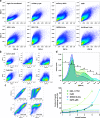
Fig. 6. Enrichment of crystal containing insect cells using fluorescence-based cell sorting.
a Forward (FSC-A) and side scatter (SSC-A) are no viable parameters for cell sorting due to a high similarity in the scattering behavior of crystal-containing and non-containing infected insect cells. b Evaluation of forward (FSC-A) and side scatter (SSC-A) against the fluorescence of EYFP does not produce a selectable population of crystal-containing cells that significantly differs between crystal-containing and non-containing cell cultures. c A high positive correlation is visible between EYFP production and cytosolic as well as endoplasmic target protein production in infected High Five cells. If the target protein is co-translated into the ER, the double negative population shifts towards a single positive population, indicating plasma membrane disruption, while the ER membranes remain intact. d FACS-based selection of infected cells based on their EYFP fluorescence allows an enrichment of crystal containing cells from the original culture. Straight lines in the lower plot indicate the fraction of crystal containing cells before cell sorting. Fractions are corresponding to the gates shown in the upper panel.
Fig. 7
Fig. 7. Intracellular crystal growth and data collection allow solving protein structures with genuine cofactors.
a A MicroMesh mount loaded with High Five cells containing IMPDH ori crystals. Both the helical line scan and resulting dozor heat map generated at the P14 beamline (PETRA III, DESY, Hamburg) are shown. b A CrystalDirectTM plate for RT diffraction data collection (asterisk) is mounted on the Arinax MD3 diffractometer in an upright position (upper panel). Below, the loaded CrystalDirectTM plate is shown with HighFive cells carrying HEX-1 cyto crystals (close-up, right panel, arrow) adherent to the bottom foil. Next to each cell-containing well is a water-filled reservoir to keep humidity high and prevent cells from drying. c Cartoon representation of the HEX-1 ori structure. d IMPDH ori structure with natural ligands (ATP and GDP) in the Bateman domain and phosphates bound in the IMP binding site of the catalytic domain (spheres). Bateman domain and Finger domain in full colors, catalytic domains in light colors. e GDP within the canonical binding site 2 of the IMPDH ori Bateman domain. The omit map calculated with simulated annealing is shown at 3.0 sigma. f ATP within the canonical binding site 1 of the IMPDH ori Bateman domain. For clarity, phosphate moieties in the alternate conformation are shown in 50% transparency. The ligand omit map is shown at 3.0 sigma.

Similar articles

Cited by

References

    1. Fan GY, et al. In vivo calcineurin crystals formed using the baculovirus expression system. Microsc. Res. Tech. 1996;34:77–86. doi: 10.1002/(SICI)1097-0029(19960501)34:1<77::AID-JEMT11>3.0.CO;2-M. - DOI - PubMed
    1. Brandariz-Nuñez A, Menaya-Vargas R, Benavente J, Martinez-Costas J. Avian reovirus microNS protein forms homo-oligomeric inclusions in a microtubule-independent fashion, which involves specific regions of its C-terminal domain. J. Virol. 2010;84:4289–301. doi: 10.1128/JVI.02534-09. - DOI - PMC - PubMed
    1. Hasegawa, H. et al. In vivo crystallization of human IgG in the endoplasmic reticulum of engineered Chinese hamster ovary (CHO.) cells. J. Biol. Chem. 286, 19917–19931 (2011). . - PMC - PubMed
    1. Doye JPK, Poon WCK. Protein crystallization in vivo. Curr. Opin. Colloid Interface Sci. 2006;11:40–46. doi: 10.1016/j.cocis.2005.10.002. - DOI
    1. Yamamoto M, et al. Protein microcrystallography using synchrotron radiation. IUCrJ. 2017;4:529–539. doi: 10.1107/S2052252517008193. - DOI - PMC - PubMed

Substances