. 2024 Feb 24;15(1):1699.
doi: 10.1038/s41467-024-45968-8.

Single-molecule RNA sizing enables quantitative analysis of alternative transcription termination

Affiliations

Single-molecule RNA sizing enables quantitative analysis of alternative transcription termination

Gerardo Patiño-Guillén et al. Nat Commun. .

Abstract

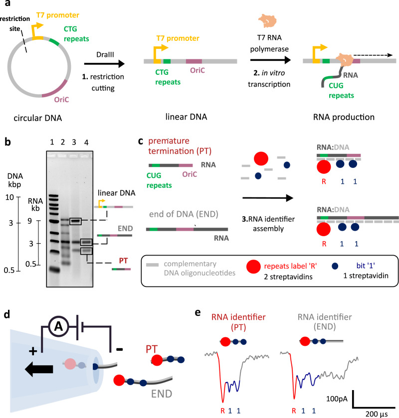
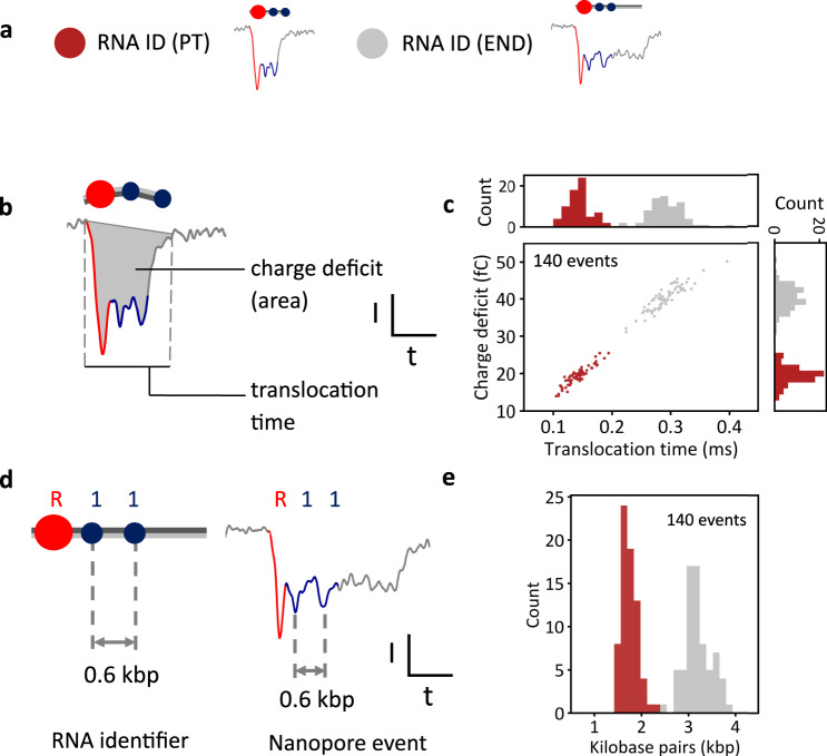
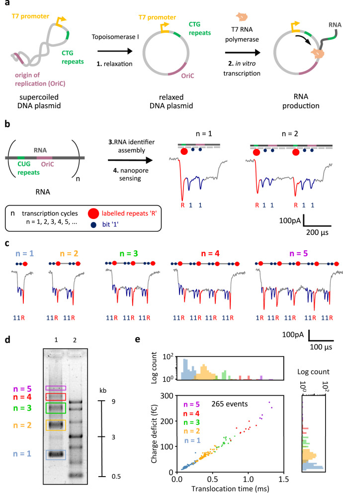
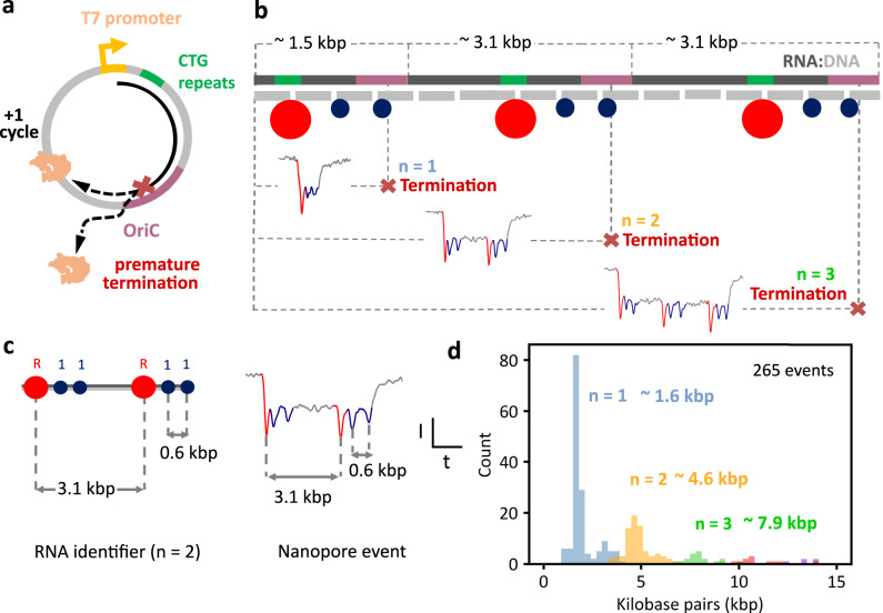
Transcription, a critical process in molecular biology, has found many applications in RNA synthesis, including mRNA vaccines and RNA therapeutics. However, current RNA characterization technologies suffer from amplification and enzymatic biases that lead to loss of native information. Here, we introduce a strategy to quantitatively study both transcription and RNA polymerase behaviour by sizing RNA with RNA nanotechnology and nanopores. To begin, we utilize T7 RNA polymerase to transcribe linear DNA lacking termination sequences. Surprisingly, we discover alternative transcription termination in the origin of replication sequence. Next, we employ circular DNA without transcription terminators to perform rolling circle transcription. This allows us to gain valuable insights into the processivity and transcription behaviour of RNA polymerase at the single-molecule level. Our work demonstrates how RNA nanotechnology and nanopores may be used in tandem for the direct and quantitative analysis of RNA transcripts. This methodology provides a promising pathway for accurate RNA structural mapping by enabling the study of full-length RNA transcripts at the single-molecule level.


Conflict of interest statement

F.B. and U.F.K. are inventors of two patents related to RNA analysis with nanopores (UK patent application no. 2113935.7, in process; UK Patent application nos. 2112088.6 and PCT/GB2022/052171, in process) submitted by Cambridge Enterprise on behalf of the University of Cambridge. U.F.K. is a co-founder of Cambridge Nucleomics. The remaining authors declare no competing interests.

Figures

Fig. 1
Fig. 1. RNA identifier (ID) assembly enables single-molecule structural analysis of RNA transcripts using nanopore sensing.
a In vitro transcription of linear DNA containing 12 CTG tandem repeats. A circular DNA construct contains a single T7RNAP promoter, the OriC, 12 CTG tandem repeats, and a DraIII restriction site. The circular DNA construct was linearized using DraIII, by cutting upstream of the T7RNAP promoter. The linear DNA was in vitro transcribed using T7RNAP. b Agarose gel electrophoresis of transcribed linear DNA indicates the presence of two RNA species (lane 4), one in which the RNA polymerase transcribes the entire linear DNA (END) and RNA of unknown nature. With nanopore sensing, it is later revealed that the lower band corresponds to RNA, where transcription is prematurely terminated within the OriC sequence (PT). Gel lanes: 1 – 1 kbp ladder; 2 – single-stranded RNA ladder; 3 – transcribed RNA from linear DNA; 4 - transcribed RNA from linear DNA treated with DNase I. Transcriptions were performed in triplicate. c RNA transcripts were hybridized with short complementary DNA oligonucleotides (~40 nt), producing RNA IDs. CUG repeats in RNA were labeled with two streptavidins (repeats label ”R”), indicating the beginning of transcription given their vicinity to the promoter. Further positioning along the transcript is achieved using bits “1” that have one streptavidin enabling their distinction from label “R”. d RNA IDs made from RNA transcripts that have different termination points (PT or END) translocating through the nanopore. Nanopore readout is based on electrophoretically driven transport of negatively charged RNA IDs through the nanopore towards a positively charged electrode. e Nanopore RNA ID readouts for PT and END RNAs are presented left and right, respectively. RNA ID for both PT and END shows downward spikes associated to the labeled repeats “R” (red) and “1” bits (blue). PT translocation finishes right after the second “1” bit hence making a clear distinction towards END translocations, which takes longer to translocate through the pore. Source data are provided as a Source Data file.
Fig. 2
Fig. 2. Quantitative analysis of single-molecule sizing of RNA determines the position of the alternative terminator.
a Example nanopore events showing translocations of RNA IDs from PT RNAs (red), where transcription terminated prematurely, and RNA IDs from long END RNAs (gray), where transcription terminated at the end of the linear DNA. b Physical parameters that are used to characterize nanopore translocation events, including event charge deficit that represents the area of an event, and translocation time. c Scatter plot of charge deficit against translocation time for RNA IDs of PT (red) and END (gray) RNA transcripts, which shows the linear dependence of both parameters. END RNA IDs require more time to translocate through nanopores. These block the ionic current for a longer time, depleting more ions, and producing a larger charge deficit. Histograms of translocation time and charge deficit show distinct distributions between both transcripts. Datapoints correspond to the translocation of RNA IDs measured within the same nanopore. The sample size was 140. d The ID design is used to convert translocation time into an estimate of the RNA length in base pairs, by knowing the base pair distance between labels and associating that distance to the time difference between the current downward spikes of the labels. e Base pair length of all molecules converted from translocation time (in c), which shows two distinct distributions. PT distribution has a mean length of (1.75 ± 0.16) kbp and END transcripts have a mean length of (3.19 ± 0.27) kbp. Errors correspond to standard deviation. Source data are provided as a Source Data file.
Fig. 3
Fig. 3. RNA identifier (ID) assembly enables single-molecule characterization of transcription of circular DNA.
a In vitro transcription of the DNA plasmid. The supercoiled plasmid contains the T7RNAP promoter, the OriC, and 12 CTG tandem repeats. The DNA was relaxed with Topoisomerase I and in vitro transcribed using T7RNAP in a rolling-circle manner. b T7RNAP produces RNA that contains multiple (“n”) copies of the plasmid sequence. Transcribed RNA was hybridized with complementary DNA oligonucleotides (~40 nt), producing RNA IDs. CUG repeats in RNA were labeled (repeats label “R”), indicating the beginning of a transcription cycle given their vicinity to the T7RNAP promoter and “1” bits were included to facilitate transcript identification. Nanopore RNA ID readouts (n = 1 and n = 2) show downward spikes associated with the labeled repeats “R” (red) and “1” bits (blue). c Nanopore readouts for RNA IDs of transcripts produced from 1 to 5 transcription cycles “n”, each showing the labeled repeats “R” and “1” bits. More example events are shown in Supplementary Figs. 12–16. RNA IDs can translocate both 5′−3′ and 3′−5′ directions through the nanopore. These events show translocations in the 3′−5′direction. Events translocating in the opposite direction are shown in Supplementary Fig. 16. d Electrophoretic mobility shift assay shows a single-stranded RNA ladder (ssRNA) on lane 2. Lane 1 shows transcription products of the relaxed plasmid, confirming nanopore readout of various transcript lengths. Gel: 1% (w/v) agarose, 1 × TBE, 0.02% sodium hypochlorite. Transcription was performed in triplicate. e Scatter plot of charge deficit against translocation time for RNA IDs of multiple transcription cycles, which shows the linear dependence of both parameters. Events with more cycles correspond to longer RNA molecules that need more time to translocate through nanopores, which blocks the ionic current for longer. Datapoints correspond to the translocation of RNA IDs measured within the same nanopore. Histograms plotted in linear scale are presented in Supplementary Fig. 17. The sample size was 265. Exemplary measurement comparing charge deficit of unfolded (linear) and folded RNA IDs is presented in Supplementary Fig. 18. Source data are provided as a Source Data file.
Fig. 4
Fig. 4. T7 RNA Polymerase’s transcription capability and premature termination analysis in circular DNA at the single-molecule level.
a Schematic of DNA plasmid, showing how termination can occur at the OriC sequence or the polymerase can continue transcribing the circular DNA for another cycle. b Schematic of RNA ID showing example events of termination after 1, 2, or 3 transcription cycles. c The ID design is used to convert translocation time into an estimate of the RNA length in base pairs. The distance between current spikes of “R” and “1” bits is translated to base pairs to obtain a conversion factor to obtain the length in base pairs for the whole event. d Histogram of normalized length (base pairs) of RNA IDs produced from the transcription of circular DNA, showing premature termination at ~1.6, ~4.6, and ~7.9 kbp in the OriC sequence, after transcription of 1 to 3 cycles, respectively. The sample size was 265 nanopore events. Raw translocation events are presented in Supplementary Figs. 19–21. Source data are provided as a Source Data file.

Similar articles

Cited by

References

    1. Engstrom MD, Pfleger BF. Transcription control engineering and applications in synthetic biology. Synth. Syst. Biotechnol. 2017;2:176–191. doi: 10.1016/j.synbio.2017.09.003. - DOI - PMC - PubMed
    1. Sousa, R. & Mukherjee, S. T7 RNA Polymerase. Prog. Nucleic Acid Res. Mol. Biol.73, 1–41 (2003). - PubMed
    1. Furukawa K, et al. Fluorescence generation from tandem repeats of a malachite green RNA aptamer using rolling circle transcription. Bioorg. Med Chem. Lett. 2008;18:4562–4565. doi: 10.1016/j.bmcl.2008.07.040. - DOI - PubMed
    1. Dang Y, Green BR. Long transcripts from dinoflagellate chloroplast minicircles suggest ‘rolling circle’ transcription. J. Biol. Chem. 2010;285:5196–5203. doi: 10.1074/jbc.M109.058545. - DOI - PMC - PubMed
    1. Jang M, Kim JH, Nam HY, Kwon IC, Ahn HJ. Design of a platform technology for systemic delivery of siRNA to tumours using rolling circle transcription. Nat. Commun. 2015;6:7930. doi: 10.1038/ncomms8930. - DOI - PMC - PubMed