. 2024 Feb 24;14(1):4534.
doi: 10.1038/s41598-024-54876-2.

Mosaic quadrivalent influenza vaccine single nanoparticle characterization

Affiliations

Mosaic quadrivalent influenza vaccine single nanoparticle characterization

Rong Sylvie Yang et al. Sci Rep. .

Abstract

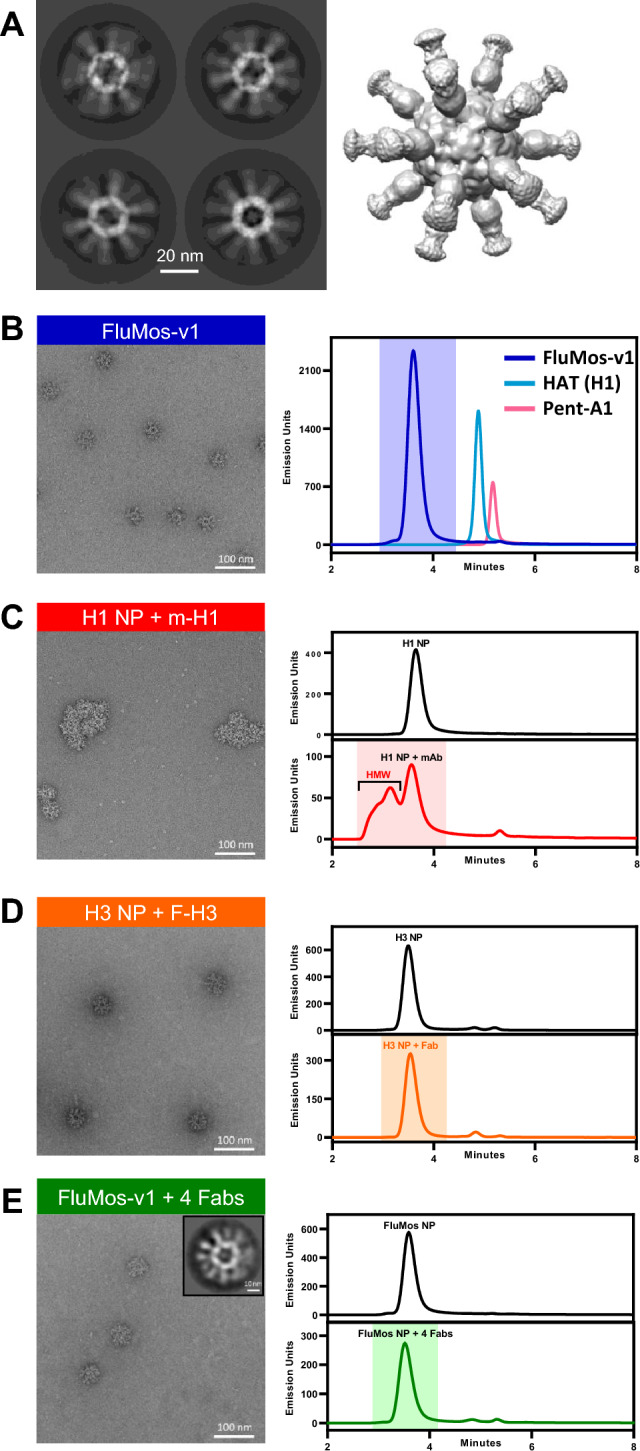
Recent work by our laboratory and others indicates that co-display of multiple antigens on protein-based nanoparticles may be key to induce cross-reactive antibodies that provide broad protection against disease. To reach the ultimate goal of a universal vaccine for seasonal influenza, a mosaic influenza nanoparticle vaccine (FluMos-v1) was developed for clinical trial (NCT04896086). FluMos-v1 is unique in that it is designed to co-display four recently circulating haemagglutinin (HA) strains; however, current vaccine analysis techniques are limited to nanoparticle population analysis, thus, are unable to determine the valency of an individual nanoparticle. For the first time, we demonstrate by total internal reflection fluorescence microscopy and supportive physical-chemical methods that the co-display of four antigens is indeed achieved in single nanoparticles. Additionally, we have determined percentages of multivalent (mosaic) nanoparticles with four, three, or two HA proteins. The integrated imaging and physicochemical methods we have developed for single nanoparticle multivalency will serve to further understand immunogenicity data from our current FluMos-v1 clinical trial.

Keywords: ELISA; Fluorescence imaging; Fluorescent labeling; Influenza vaccine; Mass spectrometry; Nanoparticle; Size-exclusion chromatography; TIRFM.


Conflict of interest statement

The authors declare no competing interests.

Figures

Figure 1
Figure 1
Overview of the structure and assembly of mosaic nanoparticle with influenza hemagglutinin (HA) antigens. (A) Imaging of FluMos-v1 using cryo-electron microscopy. Left: 2D class average images showing different views of the highly symmetrical core with long, mobile spikes (HA trimers). Right: 3D reconstruction of FluMos-v1 is shown at a low map threshold to illustrate the HA molecules on the surface of the nanoparticle. (B) Negative-stain TEM image (left) of assembled FluMos-v1 nanoparticles and SEC profiles (right) of the nanoparticles (blue) and individual components (pentamer (red), HA trimers (light blue)). (C) Negative-stain TEM image (left) and SEC profiles (right) of monovalent H1 nanoparticles. The nanoparticles were combined with the H1 strain-specific monoclonal antibody m-H1 labeled with Dylight405 (TEM and SEC profile, bottom) or analyzed without the antibody (SEC profile, top). (D) Negative-stain TEM image (left) and SEC profiles (right) of monovalent H3 nanoparticles. The nanoparticles were combined with the H3 strain-specific Fab fragment F-H3 labeled with Dylight488 (TEM and SEC bottom profile) or analyzed without the Fab (SEC top). (E) Negative-stain TEM image (left), representative 2D class average (inset), and SEC profiles (right) of quadrivalent FluMos-v1 nanoparticles. On the right, SEC profiles are the tetravalent FluMos-v1 NP (top) and FluMos-v1 mixed with labeled Fabs.
Figure 2
Figure 2
SEC Analysis of FLR-Fab Labeling of the FluMos-v1 nanoparticle. FluMos-v1 labeled with a mixture of the four Fabs (F-H1, F-HBv, F-H3, and F-HBy) (top) or the four Fab mixtures without FluMos-v1 (bottom) were injected into the SEC column. Each chromatogram represents a different fluorescent channel corresponding to the FLR-Fab-specific fluorescent label as noted above each pair or chromatograms.
Figure 3
Figure 3
SEC-FLR of DyLight-488 F-H3 and nanoparticles assembled with the H3 HA-trimer (Monovalent), the H3 and H1 HA-trimers (Bivalent), or all four HA-trimers (quadrivalent FluMos-v1). Peak area ratios are noted by the arrows and represent the fluorescence signal intensity ratio of Fab-H3 detected in monovalent (green), bivalent (red) and quadrivalent (blue) nanoparticles, with the 1:1 ratio defined as the Fab-H3 peak area of the quadrivalent nanoparticle.
Figure 4
Figure 4
FluMos-v1 nanoparticles exhibit a high degree of multivalency. (A) FluMos-v1 nanoparticles were labeled with fluorescent Fab fragments corresponding to each of the four hemagglutinin strains, then diluted in formulation buffer and imaged via TIRF microscopy. One field of view (FOV) is shown, representative of twenty random fields imaged. (B) Top row: red inset from (A) is expanded and separated by channel. Bottom row: individual nanoparticles are categorized by valency according to NIS-Elements-based analysis program. (C) Percentage of FluMos-v1 nanoparticles from all twenty FOVs in each valency category as determined by NIS-Elements-based analysis program.
Figure 5
Figure 5
Negative-stain EM of FluMos-v1, FluMos-v1 complexed with four unlabeled Fabs, and FluMos-v1 complexed with D488-F-H3. Representative micrographs are shown along with representative 2D class averages.

Similar articles

Cited by

References

    1. Kanekiyo M, Ellis D, King NP. New vaccine design and delivery technologies. J. Infect. Dis. 2019;219:S88–S96. doi: 10.1093/infdis/jiy745. - DOI - PMC - PubMed
    1. Ekiert DC, et al. Cross-neutralization of influenza A viruses mediated by a single antibody loop. Nature. 2012;489:526–532. doi: 10.1038/nature11414. - DOI - PMC - PubMed
    1. Lee PS, et al. Heterosubtypic antibody recognition of the influenza virus hemagglutinin receptor binding site enhanced by avidity. Proc. Natl. Acad. Sci. 2012;109:17040–17045. doi: 10.1073/pnas.1212371109. - DOI - PMC - PubMed
    1. Whittle JRR, et al. Broadly neutralizing human antibody that recognizes the receptor-binding pocket of influenza virus hemagglutinin. Proc. Natl. Acad. Sci. 2011;108:14216–14221. doi: 10.1073/pnas.1111497108. - DOI - PMC - PubMed
    1. Lee PS, et al. Receptor mimicry by antibody F045-092 facilitates universal binding to the H3 subtype of influenza virus. Nat. Commun. 2014;5:3614. doi: 10.1038/ncomms4614. - DOI - PMC - PubMed