. 2024 Mar 11;42(3):474-486.e12.
doi: 10.1016/j.ccell.2024.01.013. Epub 2024 Feb 22.

Chronic stress increases metastasis via neutrophil-mediated changes to the microenvironment

Affiliations

Chronic stress increases metastasis via neutrophil-mediated changes to the microenvironment

Xue-Yan He et al. Cancer Cell. .

Abstract

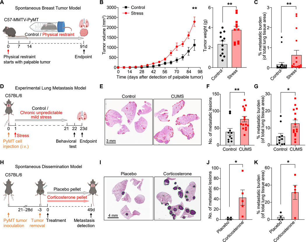
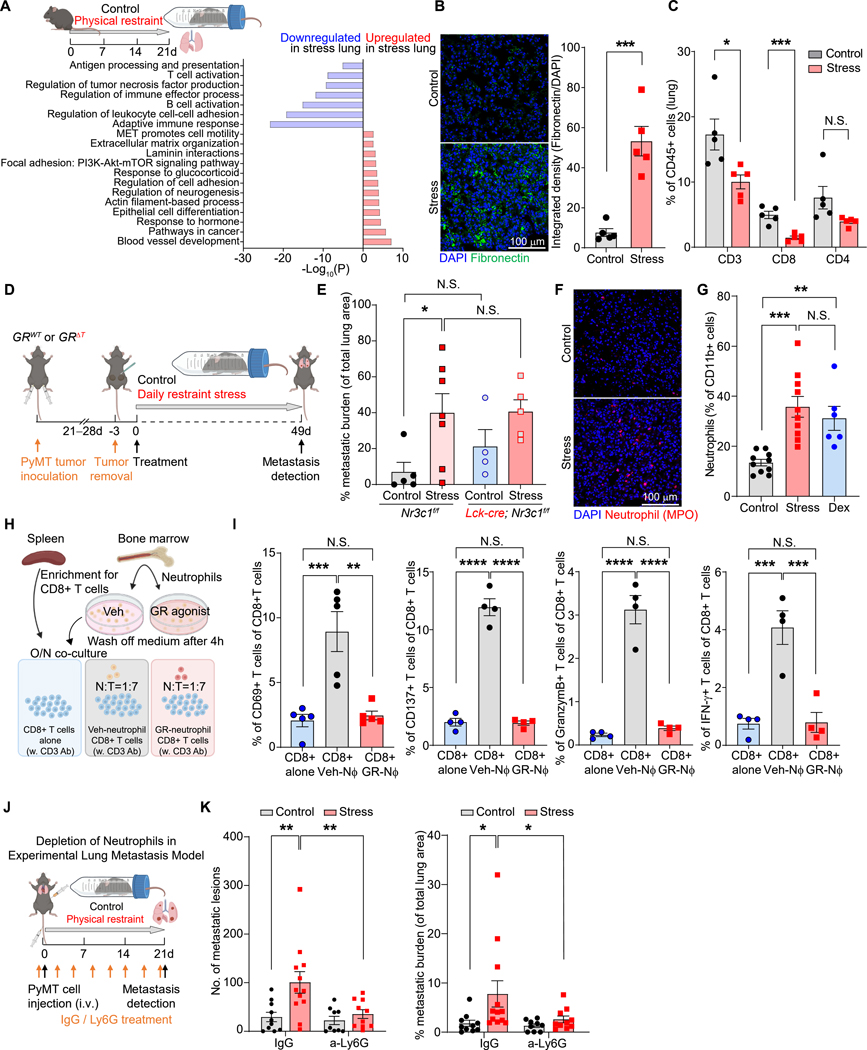
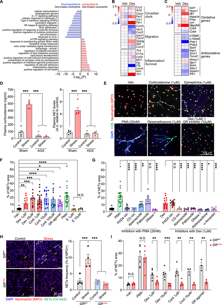
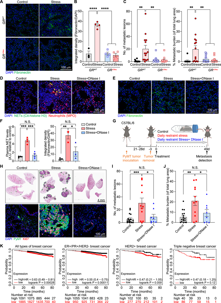
Chronic stress is associated with increased risk of metastasis and poor survival in cancer patients, yet the reasons are unclear. We show that chronic stress increases lung metastasis from disseminated cancer cells 2- to 4-fold in mice. Chronic stress significantly alters the lung microenvironment, with fibronectin accumulation, reduced T cell infiltration, and increased neutrophil infiltration. Depleting neutrophils abolishes stress-induced metastasis. Chronic stress shifts normal circadian rhythm of neutrophils and causes increased neutrophil extracellular trap (NET) formation via glucocorticoid release. In mice with neutrophil-specific glucocorticoid receptor deletion, chronic stress fails to increase NETs and metastasis. Furthermore, digesting NETs with DNase I prevents chronic stress-induced metastasis. Together, our data show that glucocorticoids released during chronic stress cause NET formation and establish a metastasis-promoting microenvironment. Therefore, NETs could be targets for preventing metastatic recurrence in cancer patients, many of whom will experience chronic stress due to their disease.

Keywords: breast cancer; chronic stress; glucocorticoids; metastasis; metastatic niche; neutrophil extracellular traps; tumor microenvironment; tumor-host interactions.


Conflict of interest statement

Declaration of interests M.E. serves on the advisory board of Cancer Cell. C.R.V. has received consulting fees from Flare Therapeutics, Roivant Sciences, and C4 Therapeutics; has served on the advisory boards of KSQ Therapeutics, Syros Pharmaceuticals, and Treeline Biosciences; and owns stock from Treeline Biosciences.

Figures

Figure 1.
Figure 1.. Chronic stress promotes metastasis
(A) Schematic of restraint stress exposure of the MMTV-PyMT breast tumor model. (B) Tumor growth curve (left) (n=10 for control, n=16 for stress) and tumor weight (right, endpoint) (n=12 for control, n=11 for stress) of primary MMTV-PyMT tumors. Tumor volume/weight indicated is the total volume/weight of all tumors for each mouse. (C) Lung metastatic burden in MMTV-PyMT mice exposed to chronic restraint stress and their controls (n=21 for control, n=16 for stress). (D) Schematic of the chronic unpredictable mild stress (CUMS) exposure of an experimental lung metastasis model. (E–G) H&E staining of lung sections after CUMS exposure at endpoint (E), number of lung metastatic lesions (F) and total metastatic burden (G) (n=10 for control, n=15 for stress). (H) Schematic of corticosterone pellet treatment in the spontaneous dissemination model. (I) Representative H&E staining of lungs at the endpoint from (H). (J, K) Number of lung metastatic lesions (J) and total metastatic burden (K) from (H) (n=5 mice/group). Data are represented as mean ± SEM. *P<0.05; **P<0.01 (B, F, G, J, K: two-tailed unpaired Student’s t-test; C: two-tailed Mann-Whitney test). See also Figures S1 and S2.
Figure 2.
Figure 2.. Chronic stress establishes a pro-metastatic lung microenvironment
(A) Gene Ontology (GO) term analysis of enriched pathways in the lungs of control and stressed mice (bulk RNA-seq; n=2 mice/condition). (B) Representative immunofluorescence staining (left) and normalized integrated density (IntDen) of fibronectin in lungs at experimental day 21. DAPI stains DNA (n=5 mice/group). (C) T cell populations in the lungs determined by flow cytometry at experimental day 21 (n=5 mice/group). (D) Schematic of chronic restraint stress exposure in the spontaneous breast cancer dissemination model used for (E–G). (E) Total metastatic burden of mice of indicated genotype at the endpoint of stress exposure (n=4–7 mice for each group). (F, G) Lung infiltration of neutrophils at day 21 (F, immunofluorescence staining for myeloperoxidase [MPO] with DAPI counterstaining; G, flow cytometry; n=10 mice in control and stress groups, n=6 in Dex group). (H) Schematic of neutrophil-CD8+ T cell co-culture assay. (I) Percentage of activated CD8+T cells (indicated by expression of CD69, CD137, Granzyme B, or IFN-γ) in neutrophil co-cultures (H) determined by flow cytometry (n=5 mice/group,). (J) Schematic of chronic restraint stress exposure in experimental lung metastasis model with neutrophil depletion. (K) Lung metastatic lesions and the total metastatic burden after stress exposure and neutrophil depletion with anti-Ly6G antibodies (n=9–12 mice/group). Data are represented as mean ± SEM. *P<0.05; **P<0.01; ***P<0.001; ****P<0.0001; N.S., not significant (B, C: two-tailed unpaired Student’s t-test; E, G, K: one-way ANOVA with Dunnett’s multiple comparisons test; I: one-way ANOVA with Tukey’s multiple comparison test). See also Figure S3.
Figure 3.
Figure 3.. Glucocorticoids induce NETs through the GR
(A) Gene Ontology (GO) term analysis of enriched pathways in bone marrow-derived neutrophils with or without dexamethasone (Dex) treatment for 4 hours (n=2 biological replicates). (B, C) Heatmap of selected genes, including (B) circadian clock genes and genes related to migration and inflammation/survival (categorized as in ) and (C) oxidative and antioxidative genes. Color scale indicates log2 fold-change in transcripts per million (TPM) for each gene relative to the average TPM of control samples (n=2 biological replicates, each pooled from two mice). (D) Enzyme-linked immunosorbent assay (ELISA) analysis of plasma samples for corticosterone (left) and NETs (right) from control and stressed mice (21 days) subjected to adrenalectomy (AGX) or sham surgery (n=4–5 mice/group). (E) NET formation assessed by immunofluorescence co-staining for anti-MPO and anti-histone H2B, with DAPI staining, of mouse neutrophils cultured overnight under indicated conditions (veh: vehicle). (F, G) NET release [quantified as % field of view (FOV) covered by NETs] of mouse neutrophils cultured under indicated conditions (dots represents FOV, neutrophils were from 2–8 mice/group). (H) Spontaneously formed NETs (yellow arrows) in ex vivo cultures of neutrophils isolated from the blood of mice of indicated genotype after 14 days of chronic restraint stress [left: representative immunofluorescence staining; right: quantification (n=4 mice/group)]. (I) Mouse neutrophils from mice of indicated genotype were cultured in vitro as indicated, and NET formation was assessed and quantified as in (E) and (F) (dots represents FOV, neutrophils were from 2 mice/group). Data are represented as mean ± SEM. *P<0.05; **P<0.01; ***P<0.001; ****P<0.0001; N.S., not significant (D, F, G, H: one-way ANOVA with Dunnett’s multiple comparisons test; I: two-tailed unpaired Student’s t-test). See also Figures S4–7.
Figure 4.
Figure 4.. Glucocorticoid-induced NETs are required for stress-induced lung metastasis
(A, B) Representative fibronectin immunofluorescence staining (A) and quantification of staining (B) in the lungs of mice with indicated genotype, subjected or not subjected to chronic restraint stress for 21 days (n=3–4 mice/group). (C) Number of lung metastatic lesions and total lung metastatic burden of mice with indicated genotype using the spontaneous dissemination model followed by chronic restraint stress for 49 days (experimental design as in Fig. 2D; n=10–14 mice/group). (D–F) Stress-induced NET formation in the lungs of mice (D), detected by immunofluorescence staining (yellow arrows indicate NETs; lungs analyzed 24 days after primary tumor resection). (E) Representative fibronectin immunofluorescence staining in the lungs of non-tumor-bearing mice, treated as indicated for 21 days. (F) NET plasma levels (left) and lung fibronectin expression (right) of mice treated as depicted in (G) (left: n=4 mice/group; right: n=3 mice/group). (G) Schematic of spontaneous dissemination model combined with chronic restraint stress and DNase I treatment. (H–J) (H) Representative H&E staining (upper row) and immunofluorescence for proliferating (Ki67+) PyMT cancer cells (bottom row) in lungs at endpoint (see G). (I) Lung metastatic lesions and (J) total metastatic burden at endpoint (see G) (n=8–17 mice/group). (K) Kaplan-Meier plots of the overall survival of breast cancer patients with high (black line) or low (red line) “chronic stress exposure gene signature” segmented by subtypes using data from the Kaplan-Meier Plotter (http://www.kmplot.com/, n is indicated in each plot, and subtypes are specified in the figure). Data are represented as mean ± SEM. *P<0.05; **P<0.01; ***P<0.001; ****P<0.0001; N.S., not significant (B: one-way ANOVA with Dunnett’s multiple comparisons test; C, F, G, I, J: one-way ANOVA with Tukey’s multiple comparison test). See also Figure S7.

Comment in

References

    1. Yaribeygi H, Panahi Y, Sahraei H, Johnston TP, and Sahebkar A. (2017). The impact of stress on body function: A review. EXCLI J 16, 1057–1072. 10.17179/excli2017-480. - DOI - PMC - PubMed
    1. McEwen BS (2007). Physiology and neurobiology of stress and adaptation: central role of the brain. Physiol Rev 87, 873–904. 10.1152/physrev.00041.2006. - DOI - PubMed
    1. Newton R. (2000). Molecular mechanisms of glucocorticoid action: what is important? Thorax 55, 603–613. DOI 10.1136/thorax.55.7.603. - DOI - PMC - PubMed
    1. Chida Y, Hamer M, Wardle J, and Steptoe A. (2008). Do stress-related psychosocial factors contribute to cancer incidence and survival? Nat Clin Pract Oncol 5, 466–475. 10.1038/ncponc1134. - DOI - PubMed
    1. Moreno-Smith M, Lutgendorf SK, and Sood AK (2010). Impact of stress on cancer metastasis. Future Oncol 6, 1863–1881. 10.2217/fon.10.142. - DOI - PMC - PubMed