. 2024 Mar 7;31(3):378-397.e12.
doi: 10.1016/j.stem.2024.01.011. Epub 2024 Feb 22.

An iron rheostat controls hematopoietic stem cell fate

Affiliations

An iron rheostat controls hematopoietic stem cell fate

Yun-Ruei Kao et al. Cell Stem Cell. .

Abstract

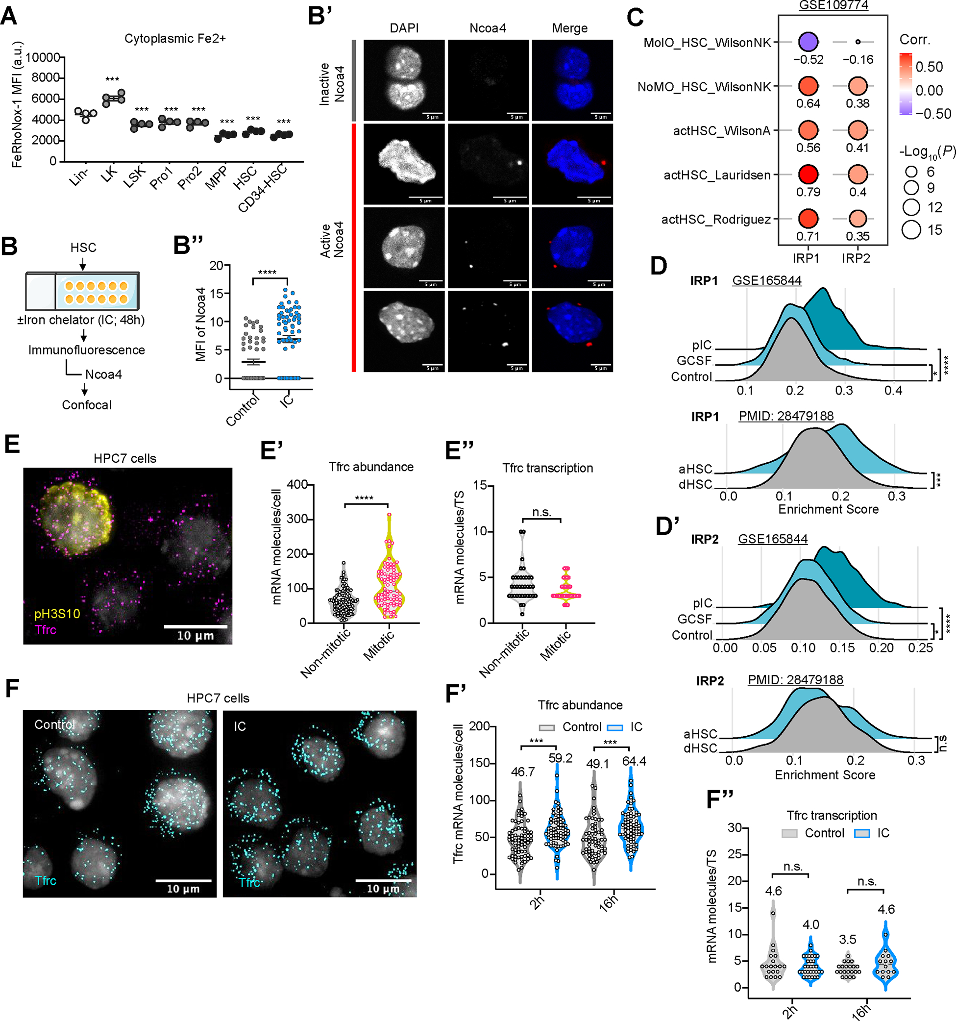
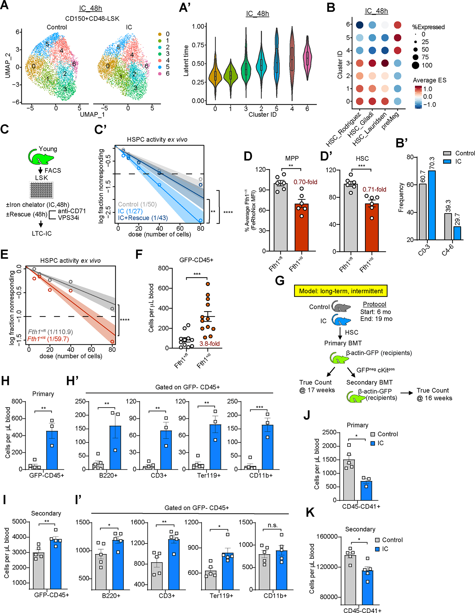
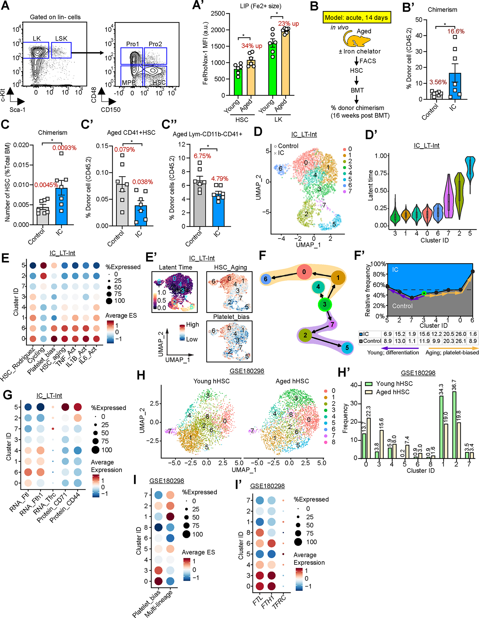
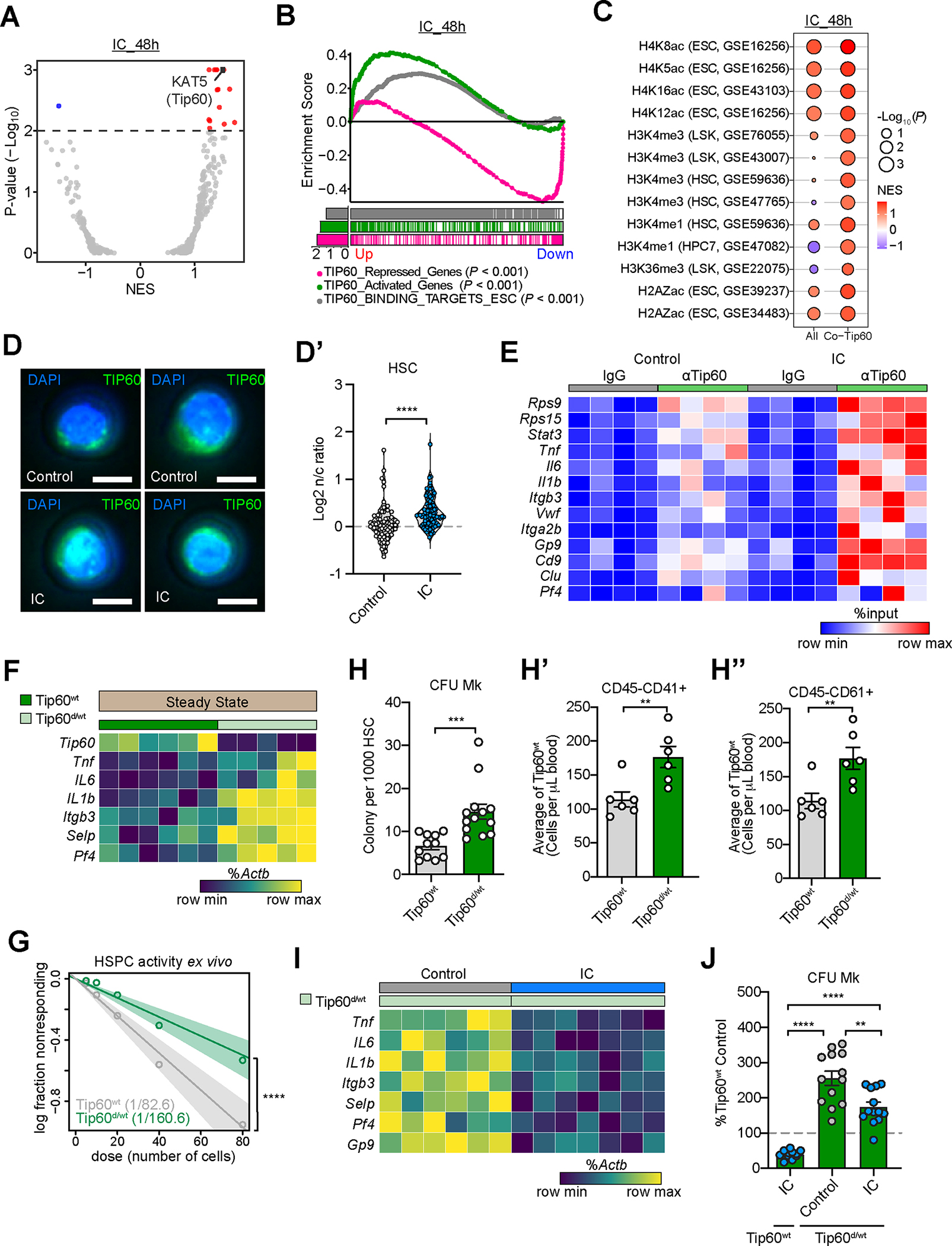
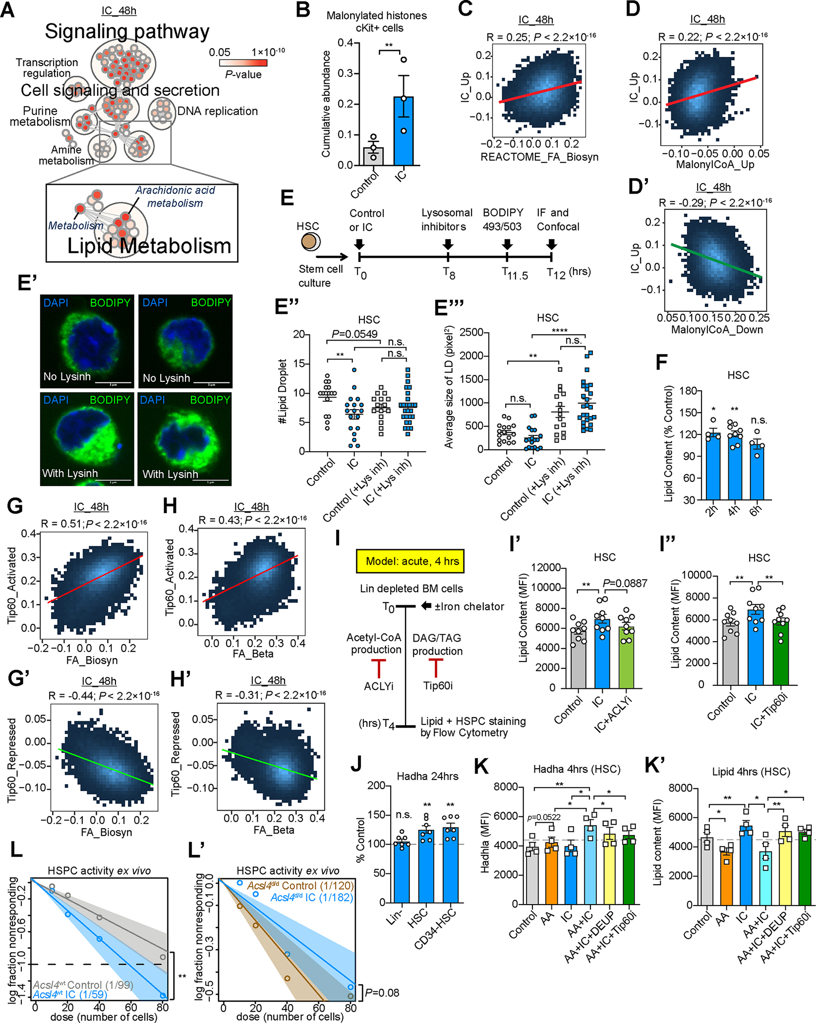
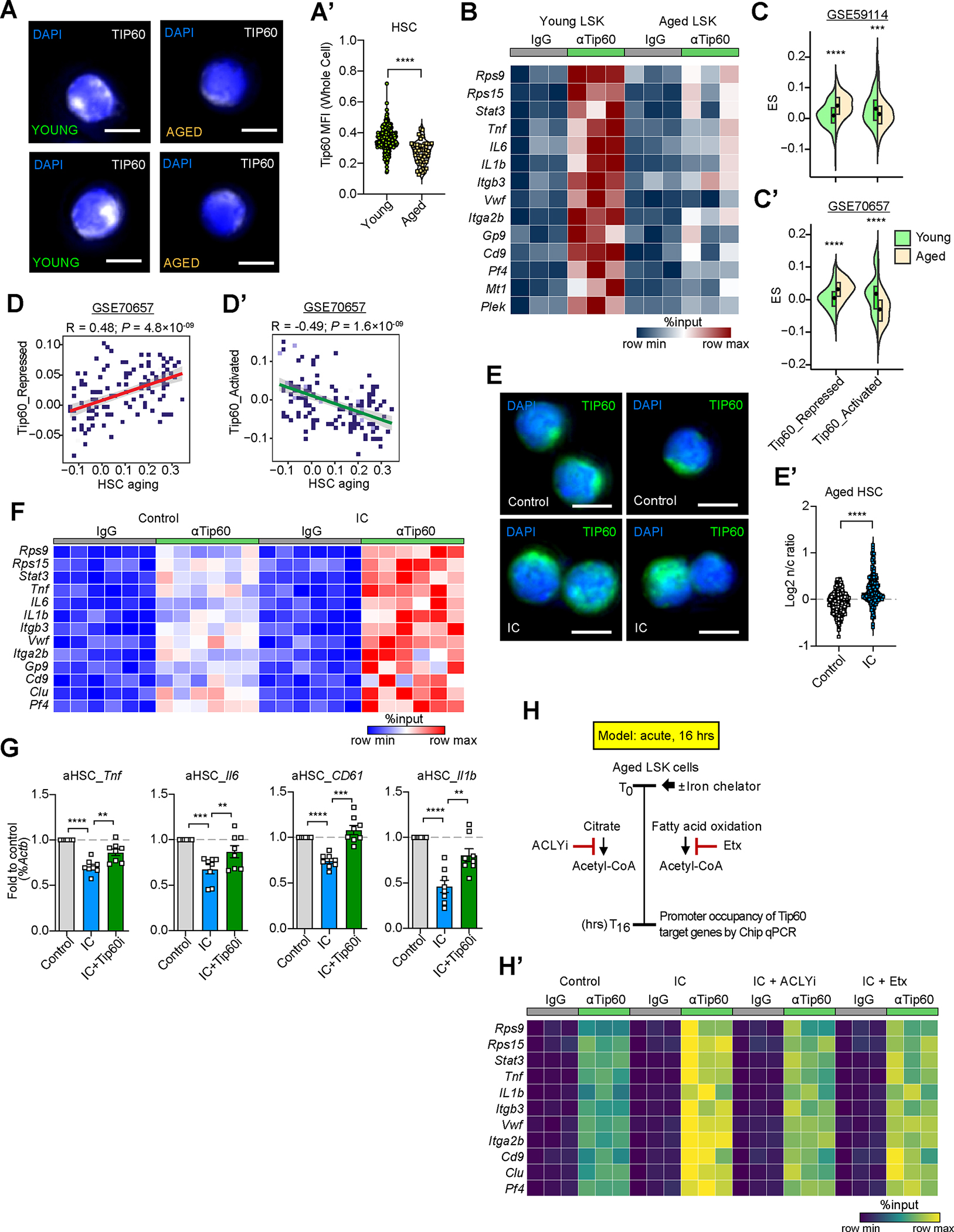
Mechanisms governing the maintenance of blood-producing hematopoietic stem and multipotent progenitor cells (HSPCs) are incompletely understood, particularly those regulating fate, ensuring long-term maintenance, and preventing aging-associated stem cell dysfunction. We uncovered a role for transitory free cytoplasmic iron as a rheostat for adult stem cell fate control. We found that HSPCs harbor comparatively small amounts of free iron and show the activation of a conserved molecular response to limited iron-particularly during mitosis. To study the functional and molecular consequences of iron restriction, we developed models allowing for transient iron bioavailability limitation and combined single-molecule RNA quantification, metabolomics, and single-cell transcriptomic analyses with functional studies. Our data reveal that the activation of the limited iron response triggers coordinated metabolic and epigenetic events, establishing stemness-conferring gene regulation. Notably, we find that aging-associated cytoplasmic iron loading reversibly attenuates iron-dependent cell fate control, explicating intervention strategies for dysfunctional aged stem cells.

Keywords: Tip60/KAT5; aging; gene regulation; hematopoiesis; iron; metabolism; stem cells.


Conflict of interest statement

Declaration of interests B.W. and U.S. have received funds for research projects and for serving on the advisory board of Novartis Pharmaceuticals.

Figures

Figure 1.
Figure 1.. LIP restriction activates the limited iron response in HSC
(A) Fe2+ LIP measurements in BM stem (HSC, CD150+CD48 LSK) and progenitor (LK, Lin c-Kit+) cells using FeRhoNox and analysis by flow cytometry. n=4. (B-B’’) Scheme of measuring ferritinophagy in HSC (B). Representative images (B’) and number of cells with activated Naco4 are shown (B’’). n=55 (Control), 66 (IC) cells. (C) Correlation of enrichment score (ES) of IRP1 and IRP2 targets with ES of signatures for activated or quiescent HSC (using GSE109774). HSC activation signatures from Rodriguez et al (PMID: 32669716), Lauridsen et al (PMID: 30021172), Wilson A et al (PMID: 19062086), no molecular overlap (NoMO) from PMID:26004780. Signatures for quiescent HSC were defined by PMID:26004780 as the molecular overlapping population (MolO). Correlation estimated using Pearson coefficient R and linear regression t-test. (D,D’) ES of IRP1 (D) and IRP2 (D’) targets in activated vs. quiescent HSC in scRNA-seq datasets GSE165844 (HSC activation after 2hr exposure to G-CSF or poly(I:C)); PMID 28479188: GFP label-retaining dormant HSC (dHSC) or GFP-negative activated HSC (aHSC). (E-E’’) Filtered and overlaid images of treated HPC7 stained with Tfrc smRNA FISH, phosphoSer10 Histone H3 (E) and DAPI. Scale bars, 10 μm; Violin plots of absolute numbers of cytoplasmic Tfrc mRNA molecules per cell (E’), pS10H3+ (mitotic; n=80) and pS10H3 (non-mitotic; n =104) cells; Violin plot of Tfrc nascent mRNA per transcription start site (TS) in non-mitotic (n=36) and mitotic (n=34) cells (E’’). (F-F’’) Filtered and overlaid images of cells stained with smRNA FISH for Tfrc, control vs. IC-treated HPC7 cells. Violin plots of Tfrc mRNA molecules per cell after 2hrs (n=71 (Control); n=74 (IC)) or 16hs (n=62 (control); n=66 (IC)) (F); or nascent Tfrc mRNA per TS after 2hrs (n=19 (Control); n=31 (IC)); or 16hrs (n=21 (control); n=14 (IC)) (F’’). If not specified otherwise, data are mean ± SEM (A, B’’). Significance indicated as *p<0.05, **p<0.01, ***p<0.001, ****p<0.0001 was calculated using Student’s t test (unpaired: B’’, D, D’, E’, E’’, F’, and F’’; paired: A). See also Figure S1 and Table S1.
Figure 2.
Figure 2.. Activation of the labile iron response enhances regenerative activity of HPSC
(A-A’) UMAP plot of cell clustering of HSC 48hrs after ex vivo treatment with DFO (IC) or vehicle control. Equal numbers of cells (4000) shown for IC and control (A). Latent time analysis of HSC clusters. Latent time of 0 represents least differentiated state (A’). (B-B’) Score of HSC signatures from Rodriguez et al. (PMID: 32669716), Giladi et al. (PMID: 29915358), and Lauridsen et al. (PMID: 30021172), as well as pre-meg (Mk-primed) signature from Rodriguez et al. (PMID: 32669716) in induced clusters (B). Proportion of HSC-like (C0, C1, C2, C3) and Mk-primed (C4, C5, C6) clusters in IC vs. control (B’). (C-C’) Experimental strategy to quantify regenerative HSC activity upon IC exposure alone or with Vps34 inhibitor and a CD71 blocking antibody (C). LTC-IC frequencies (shown in parentheses) by ELDA (C’). n=5 (D,D’) Fe2+ LIP measurements in MPP (D) and HSC (D’) cells comparing heterozygous Fth1 (Fth1+/d) or wild-type (Fth1+/fl) BM by FeRhoNox and flow cytometry. n=6 (E) Quantification of functional HSPC in heterozygous Fth1 (Fth1+/d) or wild-type (Fth1+/fl) LSK cells using LTC-IC assay. LTC-IC frequencies were shown in parentheses. n=7 (F) 6-week engraftment after intra-femural transplantation of Fth1+/d or Fth1+/fl HSC into beta-actin-GFP expressing recipients. n=11 (control) or 12 (+/d). Overall donor cell (GFPCD45+) chimerism. (G) Experimental strategy. (H-I’) Multi-lineage cell output of HSC after LT-Int IC. Donor cell engraftment of control or IC-treated HSC in recipient mice upon primary (H, H’) and secondary (I, I’) transplantation. (H,I) CD45+ donor (GFP) cells; (H’,I’) B220+ B cells, CD3+ T cells, Ter119+ erythroid cells, and CD11b+ myeloid cells. (J,K) Donor-derived cells derived platelets (GFPCD45 CD41+ FSC/SSClow) in recipient mice of primary (J) and secondary (K) transplantation. If not specified otherwise, data are mean ± SEM (D, D’, F and H-K). Significance indicated as *p<0.05, **p<0.01, ***p<0.001, ****p<0.0001 was calculated using Student’s t test (unpaired: D, D’, F, and H-K), or Poisson statistics (C’, E). See also Figure S2 and Table S2.
Figure 3.
Figure 3.. LIP restriction protects HSC regeneration during aging
(A) Gating strategy for HSPC. (A’) Fe2+ measurements in BM stem (HSC, CD150+CD48 LSK) and progenitor (LK, Linc-Kit+) cells (as in (A) from young (2–3mos.) and aged (22–24mos.) mice using FeRhoNox and FACS analysis. n 6. (B-C’’) Donor-derived cells in recipient bone marrow 16 weeks after transplantation. (B) Experimental strategy to evaluate the effect of acute iron chelation on aged HSC. (B’) total CD45.2+ cells in BM of recipients; (C) CD45.2+ HSC; (C’) CD41+ HSC; (C’’) Mk-progenitors (CD41+CD3CD4CD8B220CD11b). n=7. (D-G) CITE-seq analysis of HSPC from LT-Int mice IC at 19 mos. of age. (D) UMAP plot of HSC clustering; (D’) Latent time analysis; (E) Average signature score of different gene sets across HSC clusters. (E’) UMAP plot with scores of the latent time, signature of HSC aging and platelet bias gene sets across different clusters. (F,F’) Trajectory inference of HSC clusters determined by scVelo latent time and PAGA. Arrows indicate differentiation direction between clusters with highest connectivity (F). Relative frequency of HSC clusters in IC vs. control groups; frequencies of different clusters shown underneath (F’). Relative frequencies stacked onto a 100% scale. (G) Average abundance of Tfrc, Ftl, and Fth1, as well as CD44 and CD71 on the cell across clusters. (H-I’) Analysis of scRNA-seq data from human HSPC (GSE180298). (H) UMAP plot of clustering aged vs. young human cells. HSC cluster defined by positive expression of CD34, CRHBP, and HOPX. (H’) Proportion of clusters found altered in aged donors. (I,I’) Average signature scores of gene signatures for HSC with platelet bias or multi-lineage potential (I); average expression of FTL, FTH1, and TFRC across HSC clusters (I’). If not specified otherwise, data are mean ± SEM (A’ and B’-C’’). Significance indicated as *p<0.05 was calculated using Student’s t test (unpaired: A’, B’-C’’). See also Figure S3 and Tables S2–4.
Figure 4.
Figure 4.. LIP restriction enhances Tip60-dependent fatty acid turn-over in HSPC
(A-C) scRNA-seq of HSC following ex vivo IC (or vehicle) treatment for 48hrs. (A) Volcano plot of normalized enrichment scores (NES) of transcription factor targets (C3 GTRD transcription factor targets) by GSEAPreranked analysis. (B) GSEAPreranked analysis with Tip60-related up-regulated (Up) and down-regulated (Down) gene sets. (C) Balloon plot showing GSEAPreranked analysis of IC-associated expression profile in HSC with histone modifications and Tip60 binding-associated gene sets. (D,D’) Nuclear to cytoplasmic ratio (n/c) of Tip60 protein upon IC stimulation ex vivo for 16hrs in young HSC. Immunofluorescence images (D) and quantification (D’) are shown. n=79 or 94. Scale bars, 5μm. (E) Tip60 occupancy in gene promoters by CUT&RUN using LSK cells ex vivo cultured with IC or vehicle control for 48hrs, followed by qPCR with pull-down DNA. Data presented as % of input; color was scaled relative to the minimal (blue) and maximum (red) values of each gene. Significantly different (p<0.05) gene occupancy by Tip60 (IC vs. Control) are shown. n=4 (F) Expression of Tip60 regulated iron dependent genes in purified HSC from Tip60 wildtype (Tip60wt) and Tip60 haploinsufficient (Tip60d/wt) mice by qRT-PCR. Data are presented as % of Actb, color was scaled relative to the minimal (dark teal) and maximum (yellow) values of each gene. Significantly different (p<0.05) gene expression (wt vs. d/wt) are shown. n=6 (G) Quantification of functional HSPC in Tip60d/wt or Tip60wt LSK cells using LTC-IC assay. LTC-IC frequencies shown in parentheses. n=7 or 8 (H-H’’) Megakaryocytic output of purified HSC (CD150+CD48 LSK) fromTip60d/wt mice compared with Tip60wt controls. Ex vivo colony formation in MegaCult assays (H, n=11 or 13); in vivo platelet generation (CD45 CD41+ (H’) and CD45CD61+ (H’’); n=6) upon transplantation. (I) Expression of Tip60-regulated iron-dependent genes in activated (2-month continuous low-dose lipopolysaccharide treatment in vivo) Tip60d/wt HSC treated with vehicle or intermittent IC (LPS+IC; rescue). n=6 or 7 (J) Quantification of CFU-Mk colonies from Tip60wt or Tip60d/wt HSC, and their response to IC normalized to percent of wildtype control. n=11–13 If not specified otherwise, data are mean (H-H’’ and J) ± SEM. Significance indicated as *p<0.05, **p<0.01, ***p<0.001, ****p<0.0001 was calculated using Student’s t test (unpaired: D’ and H-H’’; paired: J), or Poisson statistics (G). See also Figure S4.
Figure 5.
Figure 5.. LIP restriction protects regenerative capacity of HSC during aging
(A) Pathway network analysis of differential genes in mouse HSC upon 48 hours IC in vitro exposure. Significantly enriched pathways (p<0.05) by NetworkAnalyst analysis. Each circle represents a pathway, the color intensity is a mapping of enrichment significance p-value. (B) Abundance of malonylated histones in control versus IC (DFO, 10 μM) treated c-Kit+ BM cells for 12hrs. n=3 (C-D’) Correlation of enrichment score of up-regulated genes in IC-treated HSC (CD150+CD48 LSK), with fatty acid biosynthesis (C) and up-regulated (D) or down-regulated (D’) DEG in malonyl-CoA treated LSK cells (GSE173256) in scRNA-seq of 48h IC-treated HSC. (E-E’’’) LD turnover in young HSC upon a 12hr IC (or vehicle) treatment. Scheme illustrating introduction of iron chelator (IC, DFO), lysosomal inhibitors (leupeptin, NH4Cl) and lipid labeling dye (BODIPY 493/503) (E). Representative immunofluorescence images (control) are shown (E’), along LD numbers (E’’) and size (E’’’). n=18–39 (F) Quantification of neutral lipid content in young HSC after acute IC for 2, 4 or 6hrs in culture using LipiGreen and analysis by flow cytometry. n=4–9 (G-G’) Correlation of enrichment scores of genes activated (G) and repressed (G’) by Tip60 and associated with FA anabolism. (H-H’) Correlation of enrichment scores of genes activated (H) and repressed (H’) by Tip60 and associated with FA catabolism. (I-I’’) Scheme illustrating quantification of neutral lipid content in young HSC upon 4hrs in culture in the presence of an iron chelator alone, in combination with ATP citrate lyase inhibition (ACLYi, 20 μM SB 204990), or a Tip60 inhibitor (TH1834, 10 μM) (I). Neutral lipid content after acute IC alone or in combination with ACLYi (I’). n=9. Lipid content after acute iron chelation alone or in combination with Tip60i (I’’). n=9. (J) Quantification of Hadha levels in Lin BM cells, HSC and CD34 HSC populations 24hrs after IC exposure compared with vehicle controls. n=7 (K-K’) Quantification of Hadha levels (K) and lipid content (K’) by flow cytometry analysis in aged HSC after a 4hr exposure to arachidonic acid (AA, 40 μM) or IC (DFO, 20μM) alone, or in combination with inhibition of lipase (DEUP, 20μM) or Tip60 (TH1834, 10 μM). n=4 (L-L’) HSPC enumeration by LTC-IC following IC treatment with LSK cells from Acsl4 wildtype (Acsl4wt, L) or Acsl4 KO (Acsl4d/d, L’). Estimated LTC-IC frequencies in parentheses. n=4 If not specified otherwise, data are mean (B, E’’, E’’’, F, I’, I’’, and J-K’) ± SEM. Significance indicated as *p<0.05, **p<0.01, ***p<0.001, ****p<0.0001 were calculated using Student’s t test (unpaired: E’’, E’’’; paired: B, F, I’, I’’, and J-K’), Poisson statistics (L and L’), or Pearson coefficient R and linear regression t test (C-D’ and G-H’). See also Figure S5 and Table S5.
Figure 6.
Figure 6.. Iron chelator-mediated labile iron pool restriction restores Tip60 activity and mitigates aging-associated HSC dysfunction
(A,A’) Tip60 protein abundance (MFI) in young and aged HSC (CD150+CD48 LSK). Immunofluorescence images of Tip60 protein abundance and subcellular localization in young and aged HSC (A). Tip60 MFI in young and aged HSC (A’). n=297 (young) and 51 (aged). Scale bars, 5μm. (B) Tip60 occupancy in gene promoters in young and aged LSK. Significantly different (p<0.05) gene occupancy by Tip60 (young LSK vs. aged LSK) is shown. n=3 (C,C’) Scores of Tip60 regulated genes in aged versus young HSC scRNAseq datasets GSE59114 (C) and GSE70657 (C’). Up-regulated (Tip60 repressed) and down-regulated (Tip60 activated) genes upon Tip60 knockout in LSK cells used as gene sets. (D,D’) Correlation of Tip60 regulated genes with HSC aging signature, using single cell expression data from GSE70657. Up-regulated (D) and down-regulated (D’) genes upon Tip60 knockout in LSK cells were used as gene sets. Correlation using Pearson coefficient R and linear regression t test. (E,E’) Nuclear to cytoplasmic ratio (n/c) of Tip60 protein in aged HSC upon IC stimulation ex vivo for 16hrs. Immunofluorescence images (E) and quantification (E’) are shown. Scale bars, 5μm. n=299 or 160. (F) Tip60 occupancy in gene promoters in aged LSK cells cultured for 48hrs with vehicle or IC. Significantly different (p<0.05) gene occupancy by Tip60 (IC vs. Control) are shown. n=6 (G) Expression changes of Tip60/Myc target genes in aged HSC after IC treatment alone or in combination with inhibition of Tip60 (Tip60i) by qPCR analysis. Fold changes of genes across treatment groups are shown. Data are mean ± SEM. n=8. (H,H’) Scheme for assessing IC-mediated Tip60 dependent acetyl-CoA production in increasing Tip60 promoter occupancy of target genes (H). (H’) Heatmap showing ChIP-qPCR of aged LSK cells ex vivo cultured for 16hrs with vehicle or IC alone, or IC in combination with inhibitors for ACLY (SB 204990, 20 μM) or CPT1a (Etomoxir, 10 μM). Significantly different (p<0.05) gene occupancy by Tip60 (IC vs. Control, ACLYi or CPT1i) are shown. n=3 Significance indicated as *p<0.05, **p<0.01, ***p<0.001, ****p<0.0001 was calculated using Student’s t test (unpaired: A’, C, C’, and E’; paired: G and H’). See also Figure S6.

Similar articles

Cited by

References

    1. Till JE, McCulloch EA, and Siminovitch L (1964). A Stochastic Model of Stem Cell Proliferation, Based on the Growth of Spleen Colony-Forming Cells. Proceedings of the National Academy of Sciences of the United States of America 51, 29–36. 10.1073/pnas.51.1.29. - DOI - PMC - PubMed
    1. Spangrude GJ, Heimfeld S, and Weissman IL (1988). Purification and characterization of mouse hematopoietic stem cells. Science 241, 58–62. 10.1126/science.2898810. - DOI - PubMed
    1. Suda T, Suda J, and Ogawa M (1983). Proliferative kinetics and differentiation of murine blast cell colonies in culture: evidence for variable G0 periods and constant doubling rates of early pluripotent hemopoietic progenitors. J Cell Physiol 117, 308–318. 10.1002/jcp.1041170305. - DOI - PubMed
    1. Baum CM, Weissman IL, Tsukamoto AS, Buckle AM, and Peault B (1992). Isolation of a candidate human hematopoietic stem-cell population. Proceedings of the National Academy of Sciences of the United States of America 89, 2804–2808. 10.1073/pnas.89.7.2804. - DOI - PMC - PubMed
    1. Morrison SJ, Wandycz AM, Akashi K, Globerson A, and Weissman IL (1996). The aging of hematopoietic stem cells. Nat Med 2, 1011–1016. 10.1038/nm0996-1011. - DOI - PubMed

Publication types

MeSH terms