. 2024 Apr 18;31(4):760-775.e17.
doi: 10.1016/j.chembiol.2024.01.010. Epub 2024 Feb 22.

Allosteric inhibition of tRNA synthetase Gln4 by N-pyrimidinyl-β-thiophenylacrylamides exerts highly selective antifungal activity

Affiliations

Allosteric inhibition of tRNA synthetase Gln4 by N-pyrimidinyl-β-thiophenylacrylamides exerts highly selective antifungal activity

Emily Puumala et al. Cell Chem Biol. .

Abstract

Candida species are among the most prevalent causes of systemic fungal infections, which account for ∼1.5 million annual fatalities. Here, we build on a compound screen that identified the molecule N-pyrimidinyl-β-thiophenylacrylamide (NP-BTA), which strongly inhibits Candida albicans growth. NP-BTA was hypothesized to target C. albicans glutaminyl-tRNA synthetase, Gln4. Here, we confirmed through in vitro amino-acylation assays NP-BTA is a potent inhibitor of Gln4, and we defined how NP-BTA arrests Gln4's transferase activity using co-crystallography. This analysis also uncovered Met496 as a critical residue for the compound's species-selective target engagement and potency. Structure-activity relationship (SAR) studies demonstrated the NP-BTA scaffold is subject to oxidative and non-oxidative metabolism, making it unsuitable for systemic administration. In a mouse dermatomycosis model, however, topical application of the compound provided significant therapeutic benefit. This work expands the repertoire of antifungal protein synthesis target mechanisms and provides a path to develop Gln4 inhibitors.

Keywords: Candida; Gln4; antifungal; fungal pathogens; glutaminyl-tRNA synthetase; translation inhibitor.


Conflict of interest statement

Declaration of interests L.E.C. and L.W. are co-founders and shareholders in Bright Angel Therapeutics, a platform company for development of novel antifungal therapeutics. L.E.C. is a science advisor for Kapoose Creek, a company that harnesses the therapeutic potential of fungi.

Figures

Fig. 1:
Fig. 1:. NP-BTA demonstrates efficacy against diverse C. albicans strains.
Two-fold dose-response assays were performed against parental (SN95, SC5314), A) azole-resistant (UPC2G648D/UPC2G648D, erg3Δ/erg3Δ, CaCi-2, CaCi-17), and B) echinocandin-resistant (FKS1/FKS1S645P, FKS1/FKS1F641S) strains of C. albicans using NP-BTA (0–100 μM), fluconazole (0–50 μg/mL), or caspofungin (0–10 μg/mL). Cultures were grown in YPD at 30 °C for 48 hours. Growth was monitored by optical density at 600 nm (OD600). Relative growth of treated samples was calculated by averaging technical duplicates and normalizing the OD600 of each treated well to the average OD600 of the untreated SC5314 wild-type control (see color bar).
Fig. 2.
Fig. 2.. NP-BTA binds C. albicans Gln4 with high affinity and inhibits tRNA aminoacylation.
A) SYPRO orange thermal shift assay reveals NP-BTA increases stability of His6-Gln4. The top panel shows the raw thermal unfolding of His6-Gln4 as an increase in the normalized relative fluorescence units (RFU) with increasing temperature. The bottom panel represents the first derivative of the raw data and was used to determine the melting temperature of His6-Gln4. B) Isothermal titration calorimetry (ITC) analysis of the interaction between His6-Gln4 (20 μM) and NP-BTA (300 μM). The top panel shows the raw differential power of each injection of NP-BTA into His6-Gln4 (dark grey) or buffer (light grey). The bottom panel shows the Wiseman isotherm of the integrated peaks fitted to a one-site binding model. The inset shows the stoichiometry (N) and thermodynamic parameters of binding. C) Raw ITC analysis of the biologically inactive NP-BTA analog STK413316 (300 μM) shows its titration into His6-Gln4 (20 μM) does not result in binding to the enzyme. D) NP-BTA inhibits His6-Gln4-catalyzed aminoacylation of tRNAGln in a dose-dependent manner. Inhibition was analyzed using a dose-response model to determine the values of IC50 and hill slope (h). All experiments were performed at least twice, with the data representing the mean ± s.e. See also Figure S1.
Fig. 3.
Fig. 3.. Co-crystal structure of NP-BTA bound to C. albicans Gln4.
A) Full-length schematic of His6-Gln4 coloured according to domain. The conserved HIGH and MSK motifs are highlighted in magenta. B) The overall structure of Gln4:NP-BTA is shown in cartoon representation covering residues 214–793 with the domains coloured using the scheme defined in panel A. Zn2+ is shown as a grey sphere and NP-BTA as red sticks. C) 2mFo-DFc map and mFo-DFc omit map are shown in blue and green mesh and contoured around the NP-BTA complex at 1.0 σ and 3.0 σ, respectively. D) Detailed surface and cartoon view of the NP-BTA binding pocket along the HIGH and MSK motifs. NP-BTA and the conserved Met496, Lys498, His265, and His268 residues are shown as sticks. The ATP binding site is outlined with a dashed line. The secondary structure is coloured according to the scheme in A. E) Sequence alignments of the HIGH and MSK motifs of representative organisms. Invariant and conserved motif residues within 4 Å of NP-BTA are highlighted in pink and grey, respectively. F) Detailed analysis of the NP-BTA binding site shown in stick representation. NP-BTA is shown in red, and the surface around it is in white. The remaining residues are coloured according to the scheme in A. See also Table S2.
Fig. 4.
Fig. 4.. Structural comparison of C. albicans Gln4:NP-BTA to E. coli GlnRS.
A) Structural superposition of the HIGH and MSK motifs of Gln4-NP-BTA with unliganded E. coli GlnRS and E. coli GlnRS in complex with 5’-O-[N-(L-glutaminyl)-sulfamoyl]adenosine (QRS) and tRNA. The comparisons are shown in two angles rotated by 180°. Conserved catalytically critical motif residues are shown as sticks. Each structure is coloured according to the legend in the bottom panel. NP-BTA and QRS are shown as red and magenta sticks, respectively. B) Schematic of the Gln4 substrate binding pocket with the ATP and Gln binding site shown in grey, the tRNA site in blue, and the MSK motif in pink. The left panel depicts the importance of Lys498 from the MSK loop in stabilizing the glutamine adenylate (Ado-Gln), based on the structure of E. coli GlnRS. The right panel depicts the putative non-competitive action of NP-BTA through binding to the MSK loop, precluding stabilization of the Ado-Gln. See also Figure S2.
Fig. 5:
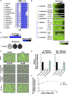
Fig. 5:. The C. albicans methionine 496 residue is important for NP-BTA activity.
A) The amino acid sequences of the conserved glutamine tRNA synthetase (GlnRS) enzyme of diverse fungal pathogens, alongside the human GlnRS, were aligned with C. albicans Gln4 using MAFFT with defaults in Jalview 2.11.2.6, with blue colour intensity corresponding to degree of conservation of each amino acid across the alignment. B) Two-fold dose-response assays were performed with NP-BTA against seven members of the Candida genus, as well as Cryptococcus neoformans, and Saccharomyces cerevisiae. Assays were completed as described in Fig. 1. C) Compound-susceptibility assays were performed against the filamentous fungi, Aspergillus fumigatus and Trichophyton rubrum. For A. fumigatus, NP-BTA activity was assessed using a two-fold dose-response assay where the fungus was incubated in RPMI at 37 °C for 48 hours with a gradient of compound. Relative viable cell number was quantified by incubating cells with Alamar Blue (1:20) after 24 hours followed by an additional 24-hour incubation at 37 °C. Fluorescence intensity at 535/595 (Ex/Em) of technical duplicates was obtained as a measure of cell viability, values were averaged, and then normalized to the untreated controls (see scale bar). For T. rubrum, NP-BTA was dissolved in potato dextrose agar (PDA) at the indicated concentrations. T. rubrum was plated and incubated for 15 days at 25 °C. Plates were captured using a ChemiDoc Imaging System. Yellow lines denote species with a Met residue aligned to C. albicans Gln4 Met496, while blue lines denote species with a Leu residue aligned to C. albicans Gln4 Met496. D) Translation inhibition assays were performed on HepG2 cells using the Click-iT HPG Alexa Fluor 488 Protein Synthesis Assay kit according to manufacturer’s instructions. HepG2 cells were seeded and treated with DMSO, NP-BTA, and control translation inhibitors (anisomycin, cycloheximide) at concentrations corresponding to the MIC80 of NP-BTA against wild-type C. albicans (6.25 μM), as well as 8-fold higher than the MIC80 concentration (50 μM). An IncuCyte live cell imager was used to image all treatment groups at 20x magnification (scale bar: 200 μm). E) The IncuCyte live cell imager’s Basic Analyzer was used for fluorescence quantification of each treatment group, relative to the DMSO control group. The green fluorescent area confluence was divided by the phase area confluence to determine the relative proportion of cells actively translating in each field. This ratio was then normalized to the relevant DMSO control and plotted. Error bars represent the mean ± SD., *** p < 0.001, n = 6. F) HEK293T-fLuc cells were seeded and treated in a two-fold dose-response assay with cycloheximide (CHX; grey) or NP-BTA (teal). Cells were treated overnight before luminescence was measured. Luminescence was normalized by averaging technical triplicate luminescence values and dividing them by the average of the untreated samples. Error bars represent the mean ± SD., n = 3. See also Figure S3.
Fig. 6:
Fig. 6:. Preliminary SAR indicates that the selected modifications to the NP-BTA scaffold do not improve bioactivity.
A) Eastern, western, and central segments of NP-BTA altered in this study. B) Activity of NP-BTA analogues against C. albicans (MIC80 values shown in μM). Analogues with eastern heterocycle modifications (shown in blue) maintain the same 3-(2-thienyl)acryloyl western elements present in NP-BTA. Similarly, analogues with modifications of the western heterocycle (shown in green) contain the N-(2-pyrimidinyl)acrylamide eastern elements. Ar = aryl; Th = thienyl. Two-fold dose-response assays were performed as described in Fig. 1. (see colour bar). C) Key interactions and hydrogen-bonds with NP-BTA in the allosteric site of Gln4. D) Summary of extra-precision docking scores (Maestro), melting temperatures (Tm, °C) resolved by thermal shift assay, and in vitro oxidative and non-ovidative metabolic stability scores expressed as percent remaining of unchanged parent compound at 30 minutes, for all molecules with bioactivity against C. albicans (MIC ≤ 100 μM), in addition to inactive control molecules 21, 25, 32, 33. See also Figure S4, Figure S5, Figure S6, and Table S1.
Fig. 7:
Fig. 7:. NP-BTA displays therapeutic potential in C. elegans candidiasis and mouse dermatomycosis models.
A) C. elegans (Au37) were infected with wild-type C. albicans (SC5314) and transferred to multi-well plates containing 0 μM, 6.25 μM and 25 μM NP-BTA. Equivalent numbers of uninfected worms were also exposed to NP-BTA as controls. Survival was measured over 48 hours in three independent experiments. Kaplan-Meier analysis is presented with the statistical significance of the difference between treatment groups determined by Log rank (Mantel-Cox) test. B) Immunosuppressed CD-1 mouse skin was infected with T. mentagrophytes conidia for 4 days before treatment with 2% NP-BTA in a cream formulation, vehicle control, or no treatment. Dosing was performed twice daily for 7 days and colony forming units (CFUs) of T. mentagrophytes per gram skin were calculated and plotted above. Unpaired t-test with Welch’s correction was used to determine statistical significance, n = 8, *** p < 0.001. Photographs of denuded mouse skin infected with T. mentagrophytes under each treatment condition (right) were taken following the complete treatment regimen. See also Figure S6.

References

    1. Fisher MC, Gurr SJ, Cuomo CA, Blehert DS, Jin H, Stukenbrock EH, Stajich JE, Kahmann R, Boone C, Denning DW, et al. (2020). Threats posed by the fungal kingdom to humans, wildlife, and agriculture. MBio 11, 1–17. 10.1128/mBio.00449-20. - DOI - PMC - PubMed
    1. Bongomin F, Gago S, Oladele R, and Denning D (2017). Global and multi-national prevalence of fungal diseases—Estimate precision. J. Fungi 3, 57. 10.3390/jof3040057. - DOI - PMC - PubMed
    1. Fisher MC, Alastruey-Izquierdo A, Berman J, Bicanic T, Bignell EM, Bowyer P, Bromley M, Brüggemann R, Garber G, Cornely OA, et al. (2022). Tackling the emerging threat of antifungal resistance to human health. Nat. Rev. Microbiol. 20, 557–571. 10.1038/s41579-022-00720-1. - DOI - PMC - PubMed
    1. Fisher MC, and Denning DW (2023). The WHO fungal priority pathogens list as a game-changer. Nat. Rev. Microbiol. 21, 211–212. 10.1038/s41579-023-00861-x. - DOI - PMC - PubMed
    1. Burki T (2023). WHO publish fungal priority pathogens list. The Lancet Microbe 4, e74. 10.1016/S2666-5247(23)00003-4. - DOI - PubMed

Substances

LinkOut - more resources