. 2024 Apr;115(4):1224-1240.
doi: 10.1111/cas.16120. Epub 2024 Feb 25.

FoxO1 promotes ovarian cancer by increasing transcription and METTL14-mediated m6A modification of SMC4

Affiliations

FoxO1 promotes ovarian cancer by increasing transcription and METTL14-mediated m6A modification of SMC4

Liping Tan et al. Cancer Sci. 2024 Apr.

Abstract

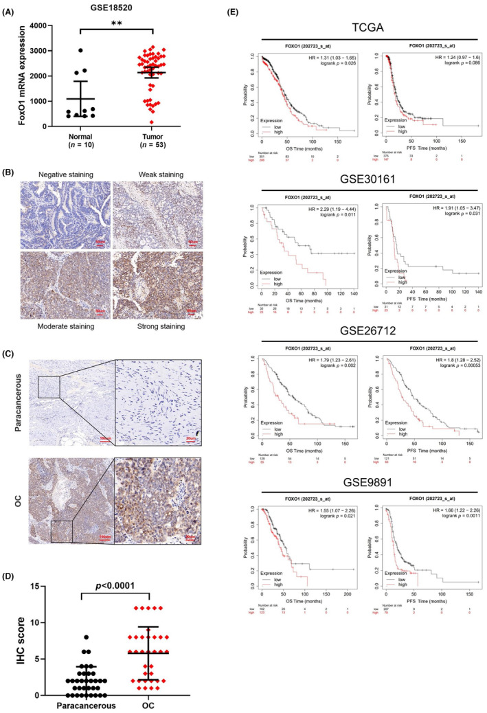
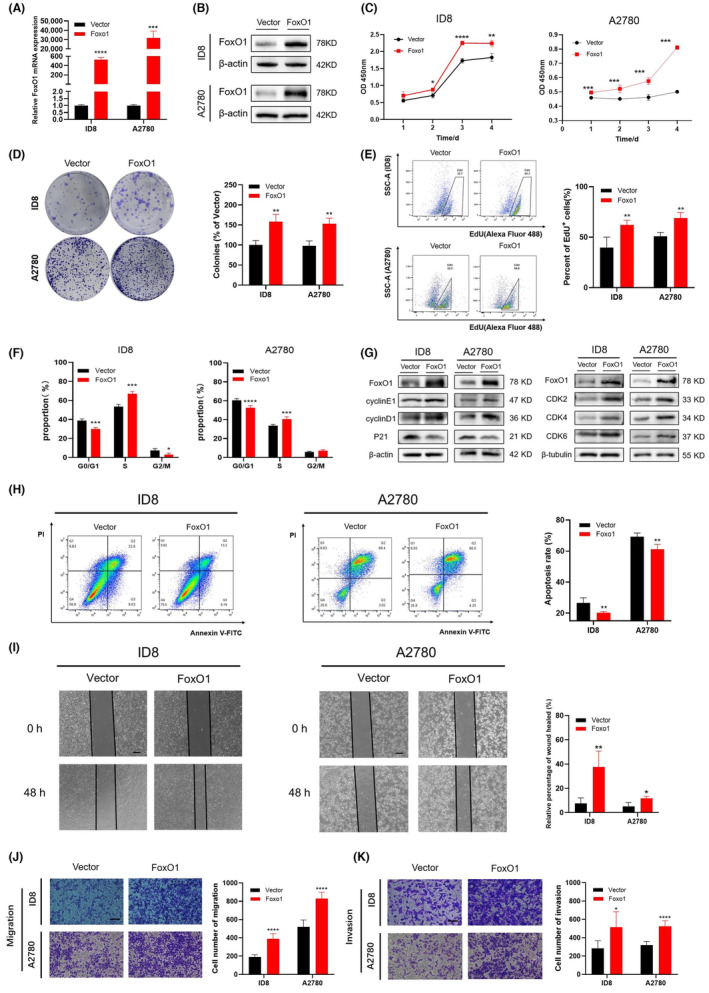
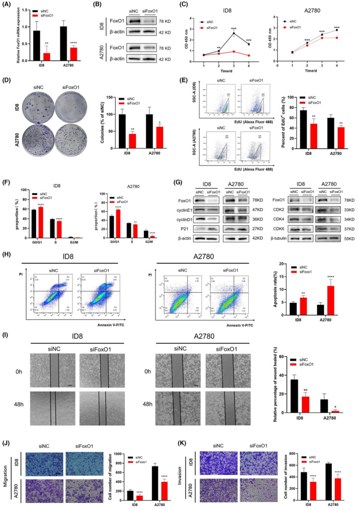
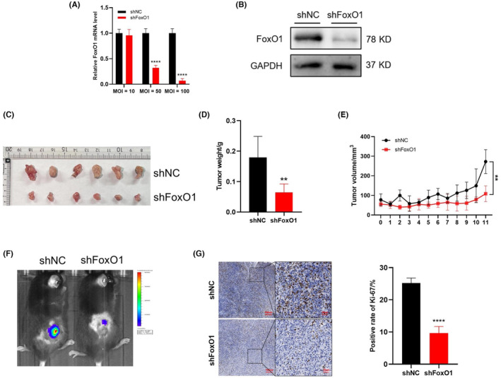
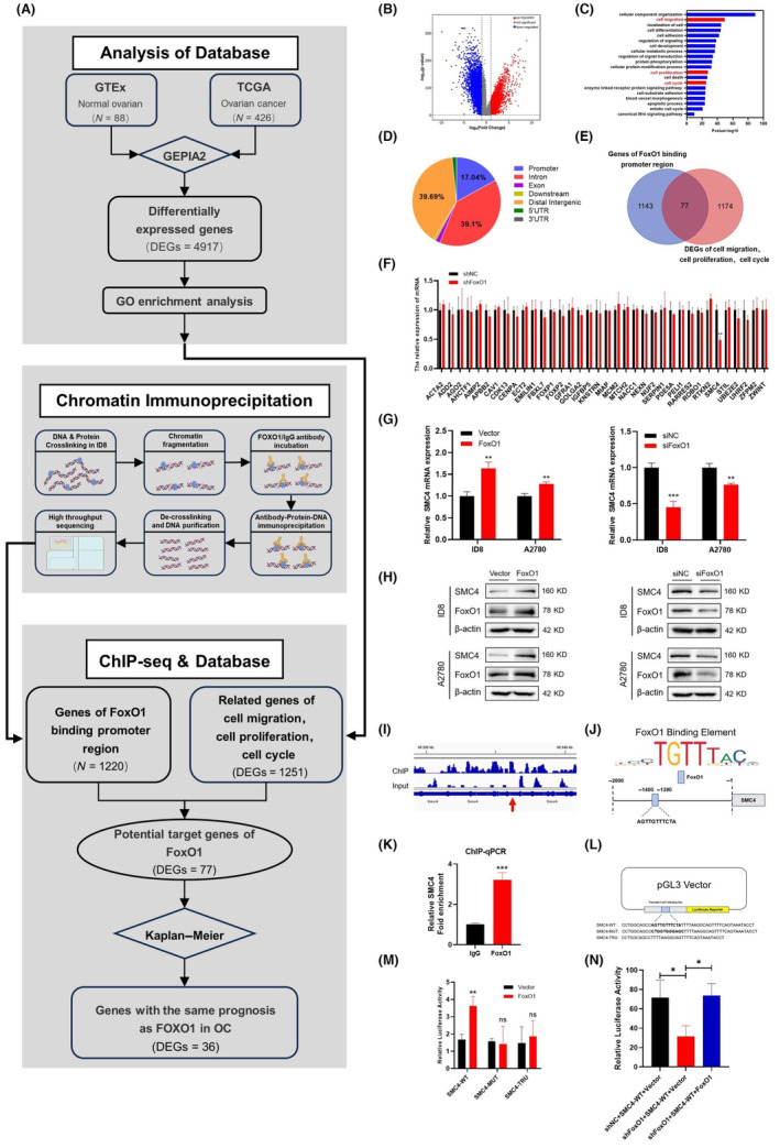
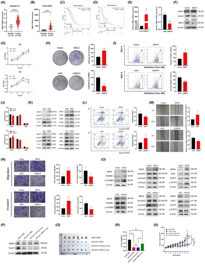
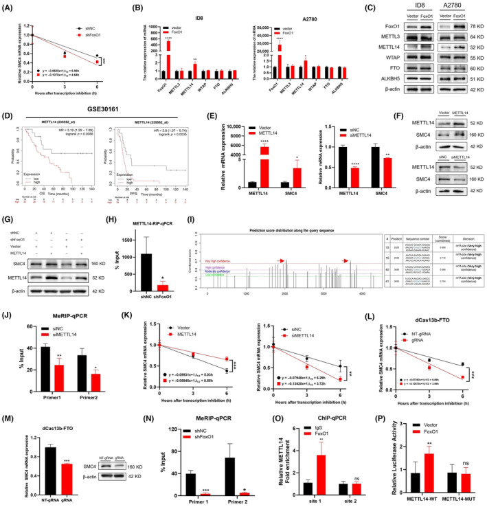
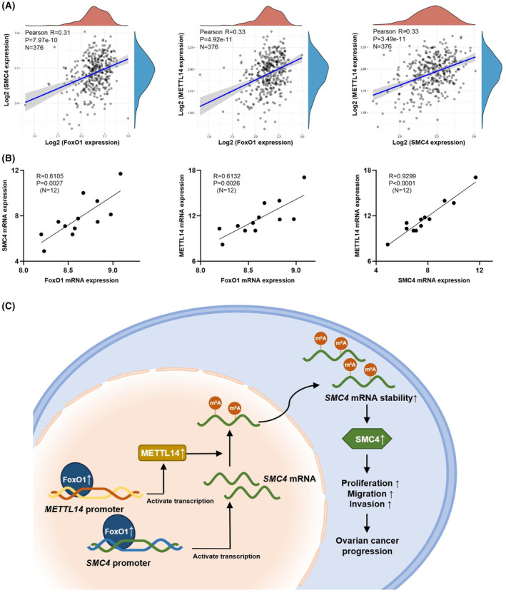
The transcription factor forkhead box protein O1 (FoxO1) is closely related to the occurrence and development of ovarian cancer (OC), however its role and molecular mechanisms remain unclear. Herein, we found that FoxO1 was highly expressed in clinical samples of OC patients and was significantly correlated with poor prognosis. FoxO1 knockdown inhibited the proliferation of OC cells in vitro and in vivo. ChIP-seq combined with GEPIA2 and Kaplan-Meier database analysis showed that structural maintenance of chromosome 4 (SMC4) is a downstream target of FoxO1, and FoxO1 promotes SMC4 transcription by binding to its -1400/-1390 bp promoter. The high expression of SMC4 significantly blocked the tumor inhibition effect of FoxO1 knockdown. Furtherly, FoxO1 increased SMC4 mRNA abundance by transcriptionally activating methyltransferase-like 14 (METTL14) and increasing SMC4 m6A methylation on its coding sequence region. The Cancer Genome Atlas dataset analysis confirmed a significant positive correlation between FoxO1, SMC4, and METTL14 expression in OC. In summary, this study revealed the molecular mechanisms of FoxO1 regulating SMC4 and established a clinical link between the expression of FoxO1/METTL14/SMC4 in the occurrence of OC, thus providing a potential diagnostic target and therapeutic strategy.

Keywords: FoxO1; METTL14; SMC4; m6A modification; ovarian cancer progression.


Conflict of interest statement

The authors declare no conflict of interest.

Figures

FIGURE 1
FIGURE 1
FoxO1 is upregulated in OC tissues, and high expression of FoxO1 is associated with a poor prognosis. (A) The expression level of the FoxO1 gene in OC and normal ovarian tissues in GSE18520. (B) The representative images of FoxO1 expression in 34 cases of OC tissues and adjacent normal control tissues detected by IHC. (C, D) Representative images of FoxO1 staining intensity (C) and quantitative analysis (D). (E) The OS and PFS of OC patients in TCGA and GEO datasets were analyzed using the Kaplan–Meier database.
FIGURE 2
FIGURE 2
Overexpression of FoxO1 promotes proliferation, migration, and invasion and inhibits apoptosis of OC cells in vitro. (A, B) Comparison of FoxO1 mRNA and protein expression in OC cell lines transfected with FoxO1 overexpression plasmid or empty vector. (C) Cell viability was analyzed using the CCK‐8 method. (D) Representative images of colonies determined by colony formation experiment. (E–H) The cell proliferation detected by EdU (E), cell cycle (F), and apoptosis (H) were determined by flow cytometry; (G) G1/S phase marker proteins were detected by western blot. (I) Scratch healing assay was used to detect cell migration. Scale bar = 400 μm. (J, K) Cell migration (J) and invasion (K) ability were detected by transwell. Scale bar = 200 μm.
FIGURE 3
FIGURE 3
Silencing FoxO1 inhibits the proliferation, migration, and invasion and promotes apoptosis of OC cells in vitro. (A, B) Comparison of FoxO1 mRNA and protein expression in OC cell lines transfected with siFoxO1 or siNC. (C) Cell viability was analyzed using the CCK‐8 assay. (D) Representative images of colonies determined by colony formation experiment. (E–H) The cell proliferation detected by EdU (E), cell cycle (F) and apoptosis (H) were determined by flow cytometry; (G) G1/S phase marker proteins were detected by western blot. (I) Scratch healing assay was used to detect cell migration. Scale bar = 400 μm. (J, K) Cell migration (J) and invasion (K) ability were detected by transwell. Scale bar = 200 μm.
FIGURE 4
FIGURE 4
FoxO1 knockdown inhibited the growth of ID8‐derived orthotopic xenografts in C57BL/6 mice. ID8 cells stably transfected with shNC or shRNA targeting FoxO1 were injected subcutaneously into the dorsal flank of C57BL/6 mice (n = 6). (A, B) Lentivirus‐generated stable FoxO1 knockdown cell lines verified by RT‐qPCR and western blot. (C) Tumor nodules, (D) tumor weight, and (E) the growth curve of tumor volume of mouse xenografts. (F) In vivo imaging of small animals to detect the fluorescence intensity of xenograft tumors. (G) The expression of Ki‐67 in shNC and shFoxO1 tumor tissues was detected by IHC.
FIGURE 5
FIGURE 5
FoxO1 directly activates SMC4 transcription in OC. (A) Screening strategy diagram of FoxO1 downstream targets in OC. (B) The volcano plot of DEGs between OC and normal ovarian tissue samples was analyzed using the GEPIA2 database. (C) Gene Ontology (GO) enrichment analysis for DEGs (top 20 are listed). (D) Distribution of FoxO1 binding sequences across the length of the DNAs in ID8 cells detected by ChIP‐seq. (E) Venn diagram of genes in the FoxO1 binding promoter region and DEGs of cell migration, proliferation, and cycle. (F) RT‐qPCR used to detect the expression of 36 potential binding targets of FoxO1 with stable knockdown of FoxO1 and its control cells. (G, H) The mRNA and protein levels of SMC4 were detected after high expression or knockdown of FoxO1. (I) IGV diagram of FoxO1 binding to the SMC4 promoter sequence. (J) The JASPAR database predicted the potential FoxO1 binding site and motif in the promoter region of SMC4. (K) ChIP‐qPCR used to determine the level of FoxO1 enrichment at the promoter of SMC4. (L) Schematic diagram of the SMC4 promoter luciferase‐reporter genes. (M) The luciferase activity with the SMC4‐WT, SMC4‐MUT or SMC4‐TRU binding site was detected after transfection of FoxO1 overexpression or control plasmid. (N) The luciferase activity with the WT binding site was detected after transfection of FoxO1 overexpression or control plasmid with stable knockdown of FoxO1 and its control ID8 cells.
FIGURE 6
FIGURE 6
SMC4 promotes the proliferation, migration, and invasion and inhibits apoptosis of ID8 cells, which is a key molecule in the process of FoxO1 promoting OC. (A, B) The expression level of SMC4 in OC tissues was compared with that in normal epithelial tissues in GEO datasets. (C, D) Kaplan–Meier database was used to analyze the OS and PFS of OC patients in TCGA and GSE9891 datasets. (E, F) The mRNA and protein levels of SMC4 after the transfection of SMC4 high‐expression plasmid or siSMC4 were detected. (G) Cell viability was analyzed using the CCK‐8 assay. (H) Representative images of colonies determined by colony formation experiment. (I–L) The cell proliferation detected by EdU (I), cell cycle (J) and apoptosis (L) were determined by flow cytometry; (K) G1/S phase marker proteins were detected by western blot. (M) Scratch healing assay used to detect cell migration. Scale bars = 400 μm. (N) Cell migration and invasion ability were detected by transwell assay. Scale bars = 200 μm. (O) The protein expression changes of TGF‐β/Smad and JAK2/STAT3 pathway molecules were detected by western blot. ID8 cells stably transfected with shNC or shRNA targeting FoxO1 were injected subcutaneously into the dorsal flank of C57BL/6 mice (n = 6), and intratumoral injection of high‐expression SMC4 lentivirus or control virus. (P) The protein expression level of SMC4 was detected in the tumor. (Q) Tumor nodules, (R) tumor weight, and (S) the growth curve of tumor volume of mouse xenografts.
FIGURE 7
FIGURE 7
FoxO1 promotes SMC4 expression by regulating METTL14‐mediated m6A modification. (A) After treatment with actinomycin D (5 μg/mL) for 0, 3, or 6 h, RT‐qPCR used to analyze the effect of low expression of FoxO1 on the half‐life of SMC4 mRNA in ID8 cells. (B) The mRNA level of m6A modification key regulatory genes was detected after high expression of FoxO1. (C) The protein level of m6A modification key regulatory genes was detected after high expression of FoxO1. (D) The Kaplan–Meier database was used to analyze the OS and PFS of OC patients in GSE30161 datasets. (E, F) Changes in mRNA and protein levels of METTL14 and SMC4 after transfection of high‐expression METTL14 plasmid or siMETTL14. (G) Western blot used to detect the effect of high expression of METTL14 after knocking down FoxO1 on the METTL14 and SMC4 protein levels. (H) METTL14‐RIP‐qPCR used to detect the enrichment of SMC4 mRNA after FoxO1 knockdown. (I) Prediction results of SMC4 mRNA in the SRAMP database show the potential sites of m6A modification. The red arrows point to sites with very high confidence. (J) MeRIP‐qPCR used to detect the enrichment of SMC4 mRNA‐predicted binding sites after low expression of METTL14. (K) RT‐qPCR to analyze the effect of high or low expression of METTL14 on the half‐life of SMC4 mRNA. (L) The half‐life of SMC4 mRNA in ID8 transfected with lentivirus containing RNA methylation editor and guide RNA (gRNA) or non‐targeting gRNA (NT‐gRNA) was detected by RT‐qPCR. (M) The expression of SMC4 mRNA and protein in ID8 transfected with lentivirus containing RNA methylation editor and gRNA or NT‐gRNA were detected. (N) MeRIP‐qPCR used to detect the enrichment of SMC4 mRNA‐predicted binding sites after FoxO1 knockdown in ID8. (O) ChIP‐qPCR used to determine the level of FoxO1 enrichment at the promoter of METTL14. (P) The luciferase activity with METTL14‐WT, METTL14‐MUT binding site was detected after transfection of FoxO1 overexpression or control plasmid.
FIGURE 8
FIGURE 8
Correlation analysis of FoxO1, SMC4 and METTL14 in OC in clinical samples and mouse models. (A) The scatter plots show the correlation of FoxO1, SMC4 and METTL14 mRNA in OC tissues of 376 OC patients from TCGA. (B) Correlation among FoxO1, SMC4 and METTL14 mRNA levels in mice xenografts (n = 12) was measured using Spearman correlation analysis. (C) Working model of SMC4 regulation by FoxO1 through transcriptional activation and increased METTL14‐mediated m6A modification to promote OC. [Correction added on 9 March 2024, after first online publication: The image for Figure 8 has been updated.]

Similar articles

Cited by

References

    1. Sung H, Ferlay J, Siegel RL, et al. Global cancer statistics 2020: GLOBOCAN estimates of incidence and mortality worldwide for 36 cancers in 185 countries. CA Cancer J Clin. 2021;71(3):209‐249. doi:10.3322/caac.21660 - DOI - PubMed
    1. Siegel RL, Miller KD, Fuchs HE, Jemal A. Cancer statistics, 2021. CA Cancer J Clin. 2021;71(1):7‐33. doi:10.3322/caac.21654 - DOI - PubMed
    1. Zhu JW, Charkhchi P, Akbari MR. Potential clinical utility of liquid biopsies in ovarian cancer. Mol Cancer. 2022;21(1):114. doi:10.1186/s12943-022-01588-8 - DOI - PMC - PubMed
    1. Berek JS, Kehoe ST, Kumar L, Friedlander M. Cancer of the ovary, fallopian tube, and peritoneum. Int J Gynaecol Obstet. 2018;143 Suppl 2:59‐78. doi:10.1002/ijgo.12614 - DOI - PubMed
    1. Barua A, Bahr JM. Ovarian cancer: applications of chickens to humans. Annu Rev Anim Biosci. 2022;10:241‐257. doi:10.1146/annurev-animal-021419-084001 - DOI - PubMed

MeSH terms