Light-controlled soft bio-microrobot
- PMID: 38403642
- PMCID: PMC10894875
- DOI: 10.1038/s41377-024-01405-5
Light-controlled soft bio-microrobot
Abstract
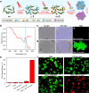
Micro/nanorobots hold exciting prospects for biomedical and even clinical applications due to their small size and high controllability. However, it is still a big challenge to maneuver micro/nanorobots into narrow spaces with high deformability and adaptability to perform complicated biomedical tasks. Here, we report a light-controlled soft bio-microrobots (called "Ebot") based on Euglena gracilis that are capable of performing multiple tasks in narrow microenvironments including intestinal mucosa with high controllability, deformability and adaptability. The motion of the Ebot can be precisely navigated via light-controlled polygonal flagellum beating. Moreover, the Ebot shows highly controlled deformability with different light illumination duration, which allows it to pass through narrow and curved microchannels with high adaptability. With these features, Ebots are able to execute multiple tasks, such as targeted drug delivery, selective removal of diseased cells in intestinal mucosa, as well as photodynamic therapy. This light-controlled Ebot provides a new bio-microrobotic tool, with many new possibilities for biomedical task execution in narrow and complicated spaces where conventional tools are difficult to access due to the lack of deformability and bio-adaptability.
© 2024. The Author(s).
Conflict of interest statement
The authors declare no competing interests.
Figures


Similar articles
-
A diatom-based biohybrid microrobot with a high drug-loading capacity and pH-sensitive drug release for target therapy.Acta Biomater. 2022 Dec;154:443-453. doi: 10.1016/j.actbio.2022.10.019. Epub 2022 Oct 13. Acta Biomater. 2022. PMID: 36243369
-
Light-deformable microrobots shape up for the biological obstacle course.Light Sci Appl. 2024 May 6;13(1):103. doi: 10.1038/s41377-024-01448-8. Light Sci Appl. 2024. PMID: 38710694 Free PMC article.
-
Light-powered phagocytic macrophage microrobot (phagobot): both in vitro and in vivo.Light Sci Appl. 2025 May 19;14(1):202. doi: 10.1038/s41377-025-01881-3. Light Sci Appl. 2025. PMID: 40383739 Free PMC article.
-
External Power-Driven Microrobotic Swarm: From Fundamental Understanding to Imaging-Guided Delivery.ACS Nano. 2021 Jan 26;15(1):149-174. doi: 10.1021/acsnano.0c07753. Epub 2021 Jan 8. ACS Nano. 2021. PMID: 33417764 Review.
-
Microrobotic Swarms for Cancer Therapy.Research (Wash D C). 2025 Apr 29;8:0686. doi: 10.34133/research.0686. eCollection 2025. Research (Wash D C). 2025. PMID: 40302783 Free PMC article. Review.
Cited by
-
Light-driven lattice soft microrobot with multimodal locomotion.Nat Commun. 2025 Aug 28;16(1):8059. doi: 10.1038/s41467-025-62676-z. Nat Commun. 2025. PMID: 40877315 Free PMC article.
-
Forward and backward control of an ultrafast millimeter-scale microrobot via vibration mode transition.Sci Adv. 2024 Oct 25;10(43):eadr1607. doi: 10.1126/sciadv.adr1607. Epub 2024 Oct 25. Sci Adv. 2024. PMID: 39453994 Free PMC article.
-
Microalgae-based hydrogel drug delivery system for treatment of gouty arthritis with alleviated colchicine side effects.Bioact Mater. 2025 May 31;52:17-35. doi: 10.1016/j.bioactmat.2025.05.021. eCollection 2025 Oct. Bioact Mater. 2025. PMID: 40519363 Free PMC article.
-
Neural stimulation and modulation with sub-cellular precision by optomechanical bio-dart.Light Sci Appl. 2024 Sep 19;13(1):258. doi: 10.1038/s41377-024-01617-9. Light Sci Appl. 2024. PMID: 39300070 Free PMC article.
-
Harnessing chemistry for plant-like machines: from soft robotics to energy harvesting in the phytosphere.Chem Commun (Camb). 2025 Apr 22;61(34):6246-6259. doi: 10.1039/d4cc06661h. Chem Commun (Camb). 2025. PMID: 40177903 Free PMC article. Review.
References
-
- Pan T, et al. Bio-micromotor tweezers for noninvasive bio-cargo delivery and precise therapy. Adv. Funct. Mater. 2022;32:2111038.
Grants and funding
- 61975065/National Natural Science Foundation of China (National Science Foundation of China)
- 12204196/National Natural Science Foundation of China (National Science Foundation of China)
- 32271405/National Natural Science Foundation of China (National Science Foundation of China)
- 61975065/National Natural Science Foundation of China (National Science Foundation of China)
- 61975065/National Natural Science Foundation of China (National Science Foundation of China)
- 61975065/National Natural Science Foundation of China (National Science Foundation of China)
- 61975065/National Natural Science Foundation of China (National Science Foundation of China)
- 61975065/National Natural Science Foundation of China (National Science Foundation of China)
- 61975065/National Natural Science Foundation of China (National Science Foundation of China)
- 61975065/National Natural Science Foundation of China (National Science Foundation of China)
LinkOut - more resources
Full Text Sources

