

The differences in the anatomy of the thoracolumbar and sacral autonomic outflow are quantitative
- PMID: 38403748
- PMCID: PMC10944453
- DOI: 10.1007/s10286-024-01023-6
The differences in the anatomy of the thoracolumbar and sacral autonomic outflow are quantitative
Abstract
Purpose: We have re-evaluated the anatomical arguments that underlie the division of the spinal visceral outflow into sympathetic and parasympathetic divisions.
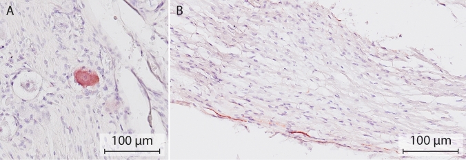
Methodology: Using a systematic literature search, we mapped the location of catecholaminergic neurons throughout the mammalian peripheral nervous system. Subsequently, a narrative method was employed to characterize segment-dependent differences in the location of preganglionic cell bodies and the composition of white and gray rami communicantes.
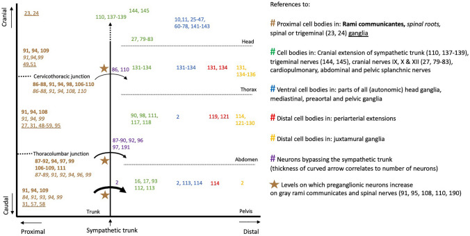
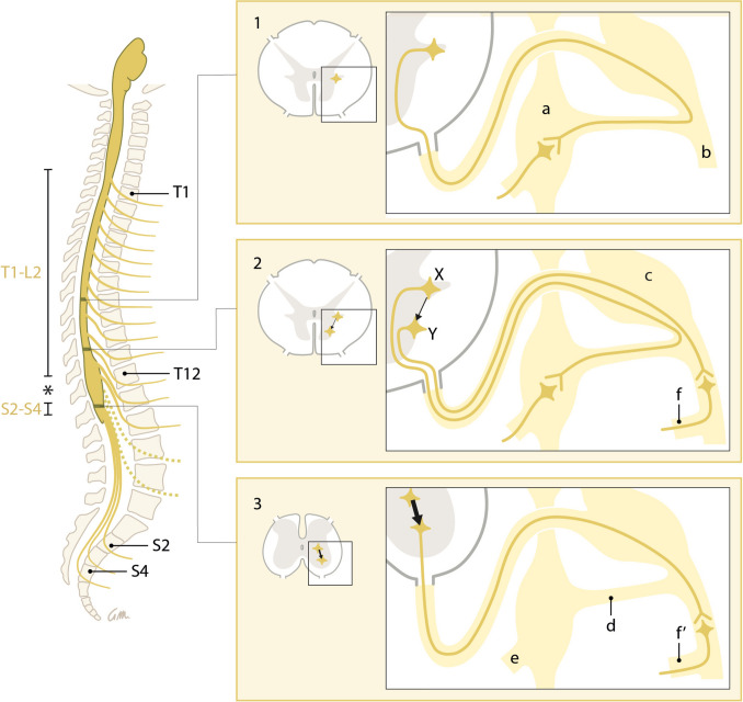
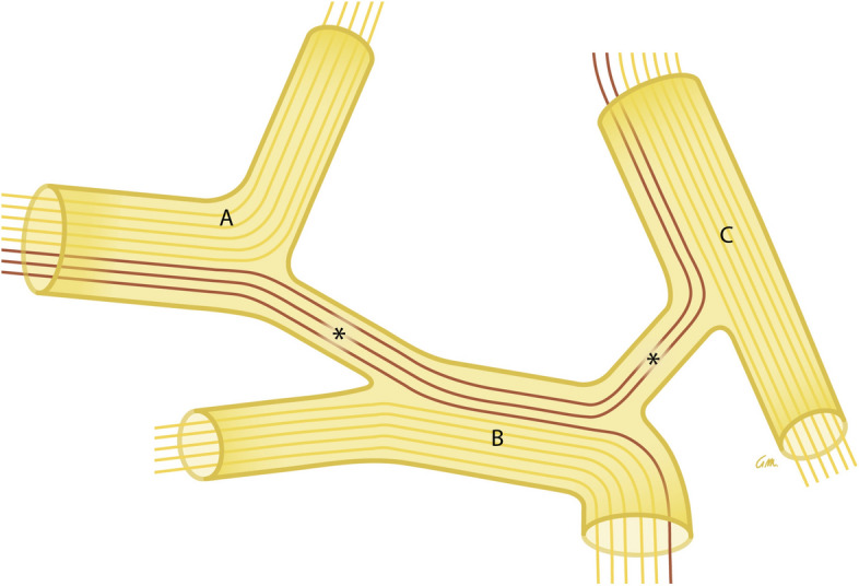
Results and conclusion: One hundred seventy studies were included in the systematic review, providing information on 389 anatomical structures. Catecholaminergic nerve fibers are present in most spinal and all cranial nerves and ganglia, including those that are known for their parasympathetic function. Along the entire spinal autonomic outflow pathways, proximal and distal catecholaminergic cell bodies are common in the head, thoracic, and abdominal and pelvic region, which invalidates the "short-versus-long preganglionic neuron" argument. Contrary to the classically confined outflow levels T1-L2 and S2-S4, preganglionic neurons have been found in the resulting lumbar gap. Preganglionic cell bodies that are located in the intermediolateral zone of the thoracolumbar spinal cord gradually nest more ventrally within the ventral motor nuclei at the lumbar and sacral levels, and their fibers bypass the white ramus communicans and sympathetic trunk to emerge directly from the spinal roots. Bypassing the sympathetic trunk, therefore, is not exclusive for the sacral outflow. We conclude that the autonomic outflow displays a conserved architecture along the entire spinal axis, and that the perceived differences in the anatomy of the autonomic thoracolumbar and sacral outflow are quantitative.
Keywords: Autonomic nervous system; Ganglion; Neural crest cells; Neuron; Postganglionic; Preganglionic.
© 2024. The Author(s).
Conflict of interest statement
The authors have no financial or non-financial conflicts of interest to declare.
Figures
Similar articles
-
The sacral autonomic outflow is sympathetic.Science. 2016 Nov 18;354(6314):893-897. doi: 10.1126/science.aah5454. Science. 2016. PMID: 27856909 Free PMC article.
-
The sympathetic and sensory components of the caudal lumbar sympathetic trunk in the cat.J Comp Neurol. 1986 Mar 1;245(1):62-73. doi: 10.1002/cne.902450105. J Comp Neurol. 1986. PMID: 3958243
-
Spinal segmental sympathetic outflow to cervical sympathetic trunk, vertebral nerve, inferior cardiac nerve and sympathetic fibres in the thoracic vagus.J Auton Nerv Syst. 1991 Mar;32(3):199-204. doi: 10.1016/0165-1838(91)90114-i. J Auton Nerv Syst. 1991. PMID: 2037767
-
Organization of lumbar spinal outflow to distal colon and pelvic organs.Physiol Rev. 1987 Oct;67(4):1332-404. doi: 10.1152/physrev.1987.67.4.1332. Physiol Rev. 1987. PMID: 2891149 Review.
-
The "sacral parasympathetic": ontogeny and anatomy of a myth.Clin Auton Res. 2018 Feb;28(1):13-21. doi: 10.1007/s10286-017-0478-7. Epub 2017 Nov 4. Clin Auton Res. 2018. PMID: 29103139 Free PMC article. Review.
Cited by
-
Anatomical Location of the Vesical Branches of the Inferior Hypogastric Plexus in Human Cadavers.Diagnostics (Basel). 2024 Apr 10;14(8):794. doi: 10.3390/diagnostics14080794. Diagnostics (Basel). 2024. PMID: 38667441 Free PMC article.
-
Gaskell, Langley, and the "para-sympathetic" idea.Elife. 2025 Mar 14;14:e104826. doi: 10.7554/eLife.104826. Elife. 2025. PMID: 40085490 Free PMC article.
-
Splanchnic neurolysis for severe cancer pain caused by abdominal paraaortic lymph node metastasis.Support Care Cancer. 2025 Feb 26;33(3):227. doi: 10.1007/s00520-025-09291-2. Support Care Cancer. 2025. PMID: 40011278
References
-
- Langley JN. The autonomic nervous system, Part I. Cambridge: W. HEFFER & SONS LTD.; 1921.
-
- Standring S Gray’s Anatomy, The Anatomical Basis of Clinical Practice. 41st Revised ed. 2016: Elsevier Health Sciences.
-
- W.F., B. and B. E.L., Medical Physiology. 3 ed. 2016: Elsevier Health Science Division.
-
- F.I.P.A.T., Terminologia Neuroanatomica 2017. - PubMed
Publication types
MeSH terms
LinkOut - more resources
Full Text Sources
Miscellaneous
