

An Splenic Artery Aneurysm and Focal Nodular Hyperplasia Associated with an Abdominal Vascular Abnormality of Hereditary Hemorrhagic Telangiectasia
- PMID: 38403756
- PMCID: PMC11557209
- DOI: 10.2169/internalmedicine.3270-23
An Splenic Artery Aneurysm and Focal Nodular Hyperplasia Associated with an Abdominal Vascular Abnormality of Hereditary Hemorrhagic Telangiectasia
Abstract
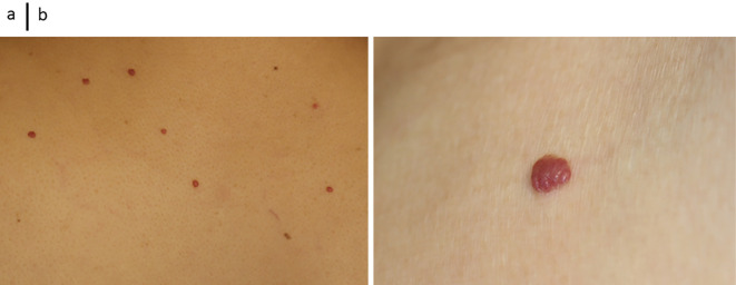
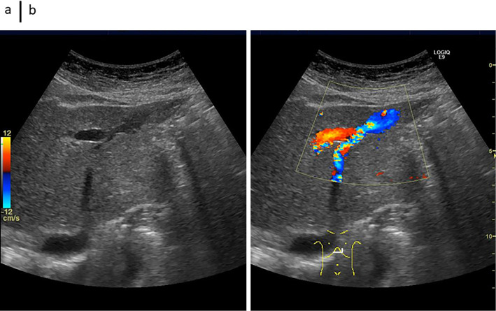
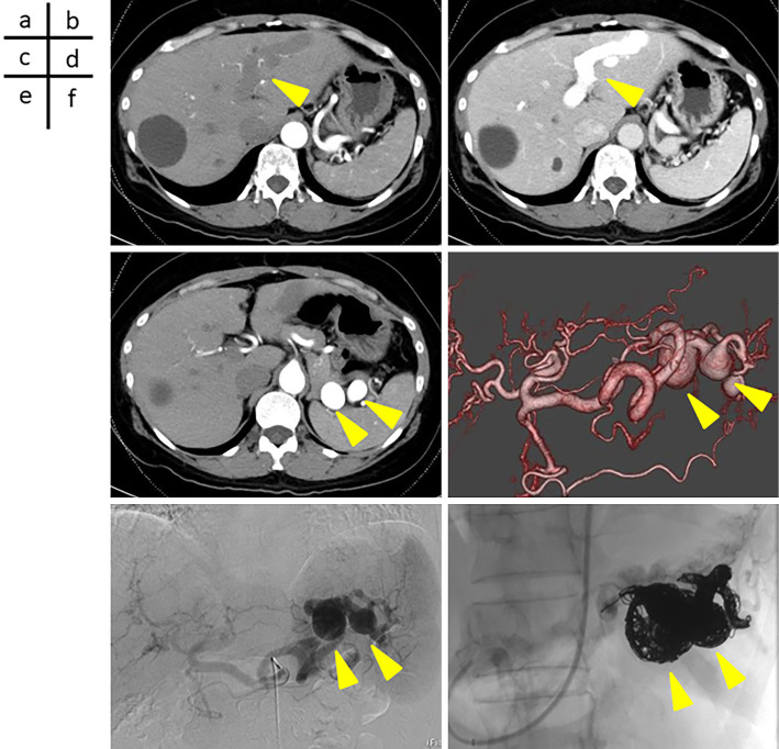
In October 2021, a 51-year-old woman developed a skin rash. Abdominal computed tomography revealed a large splenic artery aneurysm and an intrahepatic portovenous shunt. As her splenic artery aneurysm was at risk of rupture, she was referred to the Kindai University Hospital and underwent coiling surgery. In October 2023, approximately two years after she had been initially referred, contrast-enhanced ultrasound revealed findings suggestive of focal nodular hyperplasia. No reports have confirmed the occurrence of liver masses in patients with hereditary hemorrhagic telangiectasia, which is considered to be an interesting finding when investigating the mechanism of tumor development.
Keywords: Rendu-Osler-Weber disease; aneurysm; hepatic focal nodular hyperplasia; hereditary hemorrhagic telangiectasia; splenic artery; vascular malformations.
Conflict of interest statement
Figures


Similar articles
-
Hepatic involvement in hereditary hemorrhagic telangiectasia mimicking focal nodular hyperplasia.Rev Esp Enferm Dig. 2023 Mar;115(3):152-154. doi: 10.17235/reed.2022.9069/2022. Rev Esp Enferm Dig. 2023. PMID: 36043536
-
Rupture of hepatic aneurysm complicating hereditary hemorrhagic telangiectasia (Osler-Weber-Rendu disease) for which hepatic arterial coil embolization was effective.J Gastroenterol Hepatol. 2007 Dec;22(12):2352-7. doi: 10.1111/j.1440-1746.2006.03456.x. J Gastroenterol Hepatol. 2007. PMID: 18031399
-
Splenic vascular malformations and portal hypertension in hereditary hemorrhagic telangiectasia: sonographic findings.J Clin Ultrasound. 2001 Jan;29(1):56-9. doi: 10.1002/1097-0096(200101)29:1<56::aid-jcu10>3.0.co;2-o. J Clin Ultrasound. 2001. PMID: 11180187
-
Liver involvement in hereditary hemorrhagic telangiectasia (HHT).J Hepatol. 2007 Mar;46(3):499-507. doi: 10.1016/j.jhep.2006.12.008. Epub 2007 Jan 2. J Hepatol. 2007. PMID: 17239481 Review.
-
[ case of arteriovenous malformation of the pancreas with Osler-Weber-Rendu disease].Nihon Shokakibyo Gakkai Zasshi. 2008 May;105(5):719-24. Nihon Shokakibyo Gakkai Zasshi. 2008. PMID: 18460862 Review. Japanese.
References
-
- Osler W. On a family form of recurring epistaxis, associated with multiple telangiectasias of the skin and mucous membranes. Bull Johns Hopkins Hosp 12: 333-337, 1901.
-
- Hanes F. Multiple hereditary telangiectases causing hemorrhage (hereditary hemorrhage telangiectasia). Bull Johns Hopkins Hosp 20: 63-73, 1909.
-
- Shovlin CL, Guttmacher AE, Buscarini E, et al. . Diagnostic criteria for hereditary hemorrhagic telangiectasia (Rendu-Osler-Weber syndrome). Am J Med Genet 91: 66-67, 2000. - PubMed
-
- Shovlin CL, Hughes JMB, Tuddenham EGD, et al. . A gene for hereditary haemorrhagic telangiectasia maps to chromosome 9q3. Nat Genet 6: 205-209, 1994. - PubMed
Publication types
MeSH terms
LinkOut - more resources
Full Text Sources
Medical
Research Materials