Case Reports
. 2024 Nov 1;63(21):2943-2947.
doi: 10.2169/internalmedicine.3178-23. Epub 2024 Feb 26.

Pancreatic Pseudocyst after Fully Covered Self-expandable Metallic Stent Placement

Affiliations
Case Reports

Pancreatic Pseudocyst after Fully Covered Self-expandable Metallic Stent Placement

Mitsuhito Koizumi et al. Intern Med. .

Abstract

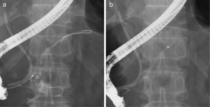
A 70-year-old woman presented with stage III pancreatic head cancer. After endoscopic sphincterotomy, a fully covered self-expandable metallic stent (FCSEMS) was placed in the common bile duct to manage jaundice. The patient developed a fever and abdominal pain 40 days after stent placement, with a suspected diagnosis of infected pancreatic pseudocyst. Purulent discharge from the papilla was observed during FCSEMS removal, and pancreatography revealed a pseudocyst connected to the main pancreatic duct. The pancreatic pseudocyst resolved after transpapillary drainage. Pancreatic pseudocysts should be suspected after biliary FCSEMS placement, and prompt removal and endoscopic drainage of the FCSEMS should be considered.

Keywords: fully covered self-expandable metallic stent; pancreatic cancer; pancreatic pseudocyst.


Conflict of interest statement

The authors state that they have no Conflict of Interest (COI).

Figures

Figure 1.
Figure 1.
a: Contrast-enhanced computed tomography revealing a mass in the pancreatic head (arrow). b: Image showing the main pancreatic duct associated with atrophy of the pancreatic body and tail (arrowhead).
Figure 2.
Figure 2.
a: Endoscopic retrograde cholangiography (ERCP) performed using pancreatic guidewire-assisted biliary cannulation. b: ERCP showing lower bile duct stricture. c: Image showing fully covered self-expandable metallic stent placement in the common bile duct after endoscopic sphincterotomy.
Figure 3.
Figure 3.
a-c: Contrast-enhanced computed tomography showing a pancreatic pseudocyst with an elongated and winding shape (arrowheads) suspected of having a connection with the main pancreatic duct (arrows) near the fully covered self-expandable metallic stent.
Figure 4.
Figure 4.
a: Purulent discharge from the papilla observed after fully covered self-expandable metallic stent removal. b: Contrast medium leakage (arrow) from the main pancreatic duct on the papillary side of the pancreatic stenosis (arrowhead) due to pancreatic head carcinoma. c: Image showing endoscopic nasopancreatic drainage (ENPD). An endoscopic biliary stent was placed in the common bile duct and an ENPD tube in the main pancreatic duct.
Figure 5.
Figure 5.
a: Image showing endoscopic biliary stent placement in the common bile duct and guidewire insertion into the main pancreatic duct and pancreatic pseudocyst. b: Image showing the placement of plastic stents into the common bile duct, main pancreatic duct, and pancreatic pseudocyst.
Figure 6.
Figure 6.
a: Computed tomography showing the endoscopic biliary and pancreatic stents and no evidence of a pancreatic pseudocyst. b: Endoscopic retrograde pancreatography showing no contrast medium leakage from the main pancreatic duct.

References

    1. Lee JH, Krishna SG, Singh A, et al. Comparison of the utility of covered metal stents versus uncovered metal stents in the management of malignant biliary strictures in 749 patients. Gastrointest Endosc 78: 312-324, 2013. - PubMed
    1. Kawakubo K, Isayama H, Nakai Y, et al. Efficacy and safety of covered self-expandable metal stents for management of distal malignant biliary obstruction due to lymph node metastases. Surg Endosc 25: 3094-3100, 2011. - PubMed
    1. Ota S, Shiomi H, Nakano R, Nishimura T, Enomoto H, Iijima H. A case of delayed pancreatic fistula after covered self-expandable metallic stent deployment for pancreatic head cancer. Clin J Gastroenterol 16: 303-309, 2023. - PubMed
    1. Rosso E, Alexakis N, Ghaneh P, et al. Pancreatic pseudocyst in chronic pancreatitis: endoscopic and surgical treatment. Dig Surg 20: 397-406, 2003. - PubMed
    1. Agalianos C, Passas I, Sideris I, Davides D, Dervenis C. Review of management options for pancreatic pseudocysts. Transl Gastroenterol Hepatol 3: 18, 2018. - PMC - PubMed

Publication types