. 2024 Feb 26;16(1):17.
doi: 10.1038/s41368-024-00286-z.

Stromal thrombospondin 1 suppresses angiogenesis in oral submucous fibrosis

Affiliations

Stromal thrombospondin 1 suppresses angiogenesis in oral submucous fibrosis

Xiao Yang et al. Int J Oral Sci. .

Abstract

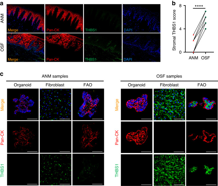
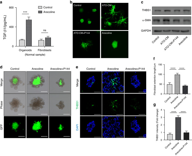
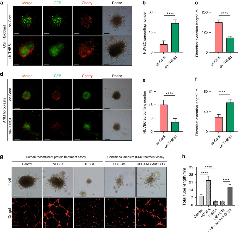
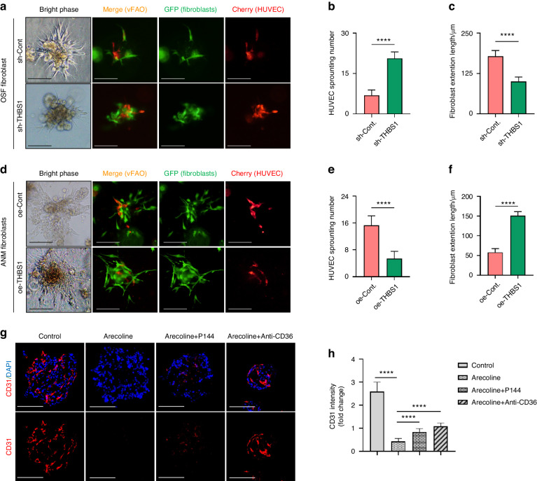
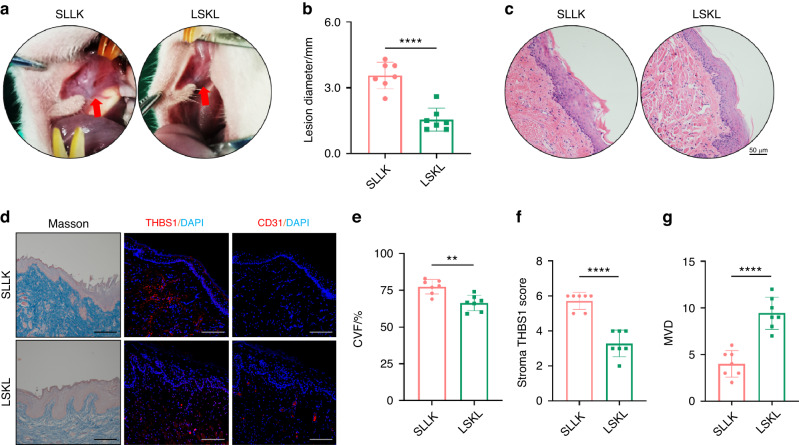
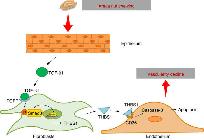
A decline in mucosal vascularity is a histological hallmark of oral submucous fibrosis (OSF), a premalignant disease that is largely induced by betel quid chewing. However, the lack of available models has challenged studies of angiogenesis in OSF. Here, we found that the expression of thrombospondin 1 (THBS1), an endogenous angiostatic protein, was elevated in the stroma of tissues with OSF. Using a fibroblast-attached organoid (FAO) model, the overexpression of THBS1 in OSF was stably recapitulated in vitro. In the FAO model, treatment with arecoline, a major pathogenic component in areca nuts, enhanced the secretion of transforming growth factor (TGF)-β1 by epithelial cells, which then promoted the expression of THBS1 in fibroblasts. Furthermore, human umbilical vein endothelial cells (HUVECs) were incorporated into the FAO to mimic the vascularized component. Overexpression of THBS1 in fibroblasts drastically suppressed the sprouting ability of endothelial cells in vascularized FAOs (vFAOs). Consistently, treatment with arecoline reduced the expression of CD31 in vFAOs, and this effect was attenuated when the endothelial cells were preincubated with neutralizing antibody of CD36, a receptor of THBS1. Finally, in an arecoline-induced rat OSF model, THBS1 inhibition alleviated collagen deposition and the decline in vascularity in vivo. Overall, we exploited an assembled organoid model to study OSF pathogenesis and provide a rationale for targeting THBS1.


Conflict of interest statement

The authors declare no competing interests.

Figures

Fig. 1
Fig. 1
Overexpression of THBS1 in OSF is recapitulated in a human FAO model. a Representative images of immunofluorescence (IF) staining showed the expression pattern of THBS1 and pan-cytokeratin (pan-CK) in OSF and normal mucosa (NM) tissue. scale bars, 100 μm. b Quantification analysis of IF staining showed the stromal THBS1 score in OSF and normal mucosa (NM) tissue. n = 10. ****P < 0.000 1. c Representative images showed the IF staining of THBS1 and pan-CK in organoid, fibroblast, and FAO generated using OSF and normal samples, scale bars, 100 μm
Fig. 2
Fig. 2
Arecoline promotes the expression of stromal THBS1 in FAO. a Enzyme-linked immunosorbent assay (ELISA) detected TGF-β1 concentration in epithelial organoids or fibroblasts derived conditional medium (CM) upon arecoline treatment (10 μg/mL). ***P < 0.001. b Representative images showed the morphology of fibroblasts cultured in the matrigel, treated with arecoline-treated organoid derived CM (ATO-CM), ATO-CM + P144 (100 μg/mL) and arecoline (10 μg/mL), scale bars, 100 μm. c IB assay showed the expression of THBS1 in fibroblasts treated ATO-CM, ATO-CM + P144 (100 μg/mL) and arecoline (10 μg/mL). d Representative images show the morphology of FAO upon treatment with arecoline (10 μg/mL) and arecoline + P144 (100 μg/mL), scale bars, 100 μm. e Representative IF staining images showed the expression pattern of THBS1 in FAO upon treatment with arecoline (10 μg/mL) and arecoline (10 μg/mL) + P144 (100 μg/mL), scale bars, 100 μm. f Fibroblast (GFP-labelled) extension length in FAO was used as a quantity index. ****P < 0.000 1. g Quantification analysis of IF staining showed the expression of THBS1 in FAO. ****P < 0.000 1
Fig. 3
Fig. 3
Fibroblast-derived THBS1 suppresses the sprouting ability of endothelial cells. af Representative images and quantitative analysis showed the mixture of fibroblasts and human umbilical vein endothelial cells (HUVECs) in the fibroblast-HUVEC clusters. Quantification analysis validated that knockdown of THBS1 in fibroblasts in OSF (OSF fibroblast) increased the sprouting number of HUVECs (b) but decreased the length of fibroblasts (c). Overexpression of THBS1 in normal fibroblasts (ANM fibroblast) reduced the sprouting number of HUVECs (d, e) but induced the length of fibroblasts (d, f), scale bars, 100 μm. ****P < 0.000 1. g Representative images of HUVEC colony in gel and the tube formation on gel, treated with vascular endothelial growth factor A (VEGFA, 1 μg/mL), THBS1 (1 μg/mL), OSF-fibroblast derived CM (OSF-CM) and OSF-CM + Anti-CD36, scale bars, 100 μm. h Quantitative analysis of the length of tubes formed by HUVECs. ****P < 0.000 1
Fig. 4
Fig. 4
Arecoline reduces the expression of endothelial marker in vascularized FAOs. af Representative images showed the distribution and morphology of fibroblasts (GFP-labeled) and HUVECs (mCherry-labeled) in the vascularized FAO (v-FAO). Quantification analysis validated that knockdown of THBS1 in fibroblasts in OSF (OSF fibroblast) increased the sprouting number of HUVECs (b) but decreased the extension length of fibroblasts (c). Overexpression of THBS1 in adjacent normal fibroblasts (ANM fibroblast) reduced the sprouting number of HUVECs (e) but induced the extension length of fibroblasts (f), scale bars, 100 μm. ****P < 0.000 1. g Representative images of IF staining showed the expression of CD31 in v-FAO treated with arecoline (10 μg/mL), arecoline (10 μg/mL) + P144 (100 μg/mL) and arecoline (10 μg/mL) + Anti-CD36. h Quantification analysis of CD31 expression in v-FAO. ****P < 0.000 1
Fig. 5
Fig. 5
Inhibiting THBS1 alleviates disease progression in a rat OSF model. a Representative image showed the white patch in oral mucosa of SD rats treated with SLLK or LSKL. b Lesion diameter was measured and quantitative analysis showed that it was much longer in SLLK group compared than that in LSKL group. n = 7. ****P < 0.000 1. c HE staining confirmed histological change of specimens in SLLK group and LSKL group, scale bars, 50 μm. d Representative images of Masson staining and IF staining showed the collagen deposition and the expression of THBS1 and CD31 in each group, scale bars, 100 μm. eg quantitative analysis of collagen volume fraction (CVF), the stromal score of THBS1 and the microvessel density (MVD) in SLLK group and LSKL group. **P < 0.01. ****P < 0.000 1
Fig. 6
Fig. 6
Schematic of the proposed mechanism by which THBS1 suppresses angiogenesis in OSF

Similar articles

Cited by

References

    1. Tilakaratne WM, Klinikowski MF, Saku T, Peters TJ, Warnakulasuriya S. Oral submucous fibrosis: review on aetiology and pathogenesis. Oral Oncol. 2006;42:561–568. - PubMed
    1. Shih YH, Wang TH, Shieh TM, Tseng YH. Oral submucous fibrosis: a review on etiopathogenesis, diagnosis, and therapy. Int. J. Mol. Sci. 2019;20:2940. - PMC - PubMed
    1. Ray JG, Chatterjee R, Chaudhuri K. Oral submucous fibrosis: a global challenge. rising incidence, risk factors, management, and research priorities. Periodontol 2000. 2019;80:200–212. - PubMed
    1. Sharma M, Radhakrishnan R. Limited mouth opening in oral submucous fibrosis: reasons, ramifications, and remedies. J. Oral Pathol. Med. 2017;46:424–430. - PubMed
    1. Qin X, Ning Y, Zhou L, Zhu Y. Oral submucous fibrosis: etiological mechanism, malignant transformation, therapeutic approaches and targets. Int. J. Mol. Sci. 2023;24:4992. - PMC - PubMed

Publication types