An atlas of gross and histologic lesions and immunohistochemical immunoreactivity during the temporal progression of aerosolized Lassa virus induced hemorrhagic fever in cynomolgus macaques
- PMID: 38404292
- PMCID: PMC10884106
- DOI: 10.3389/fcimb.2024.1341891
An atlas of gross and histologic lesions and immunohistochemical immunoreactivity during the temporal progression of aerosolized Lassa virus induced hemorrhagic fever in cynomolgus macaques
Abstract
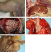
Lassa virus (LASV) causes an acute multisystemic hemorrhagic fever in humans known as Lassa fever, which is endemic in several African countries. This manuscript focuses on the progression of disease in cynomolgus macaques challenged with aerosolized LASV and serially sampled for the development and progression of gross and histopathologic lesions. Gross lesions were first noted in tissues on day 6 and persisted throughout day 12. Viremia and histologic lesions were first noted on day 6 commencing with the pulmonary system and hemolymphatic system and progressing at later time points to include all systems. Immunoreactivity to LASV antigen was first observed in the lungs of one macaque on day 3 and appeared localized to macrophages with an increase at later time points to include immunoreactivity in all organ systems. Additionally, this manuscript will serve as a detailed atlas of histopathologic lesions and disease progression for comparison to other animal models of aerosolized Arenaviral disease.
Keywords: Lassa fever; Lassa virus; cynomolgus macaques; histologic lesion; immunohistochemistry; non-human primate.
Copyright © 2024 Bohler, Cashman, Wilkinson, Johnson, Rosenke, Shamblin, Hensley, Honko and Shaia.
Conflict of interest statement
The authors declare that the research was conducted in the absence of any commercial or financial relationships that could be construed as a potential conflict of interest.
Figures


Similar articles
-
Immune-Mediated Systemic Vasculitis as the Proposed Cause of Sudden-Onset Sensorineural Hearing Loss following Lassa Virus Exposure in Cynomolgus Macaques.mBio. 2018 Oct 30;9(5):e01896-18. doi: 10.1128/mBio.01896-18. mBio. 2018. PMID: 30377282 Free PMC article.
-
Temporal Progression of Lesions in Guinea Pigs Infected With Lassa Virus.Vet Pathol. 2017 May;54(3):549-562. doi: 10.1177/0300985816677153. Epub 2016 Jan 1. Vet Pathol. 2017. PMID: 28438110
-
Pathogenesis of Lassa fever in cynomolgus macaques.Virol J. 2011 May 6;8:205. doi: 10.1186/1743-422X-8-205. Virol J. 2011. PMID: 21548931 Free PMC article.
-
Antibody therapy for Lassa fever.Curr Opin Virol. 2019 Aug;37:97-104. doi: 10.1016/j.coviro.2019.07.003. Epub 2019 Aug 8. Curr Opin Virol. 2019. PMID: 31401518 Review.
-
Current research for a vaccine against Lassa hemorrhagic fever virus.Drug Des Devel Ther. 2018 Aug 14;12:2519-2527. doi: 10.2147/DDDT.S147276. eCollection 2018. Drug Des Devel Ther. 2018. PMID: 30147299 Free PMC article. Review.
References
-
- Bonwitt J., Sáez A. M., Lamin J., Ansumana R., Dawson M., Buanie J., et al. . (2017). At home with mastomys and rattus: human-rodent interactions and potential for primary transmission of lassa virus in domestic spaces. Am. J. Trop. Med. Hygiene 96 (4), 935–943. doi: 10.4269/ajtmh.16-0675 - DOI - PMC - PubMed
MeSH terms
Substances
LinkOut - more resources
Full Text Sources

