

Laser speckle imaging of the hippocampus
- PMID: 38404300
- PMCID: PMC10890870
- DOI: 10.1364/BOE.507371
Laser speckle imaging of the hippocampus
Abstract
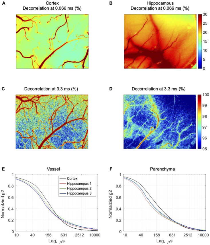
Research on hippocampal blood flow is essential for gaining insight into its involvement in learning and memory and its role in age-related cognitive impairment and dementia. In this study, we applied laser speckle contrast imaging (LSCI) and dynamic light scattering imaging (DLSI) to monitor perfusion in mouse hippocampus via a chronic, optically transparent window. LSCI scans showed hippocampal blood vessels appear more out of focus than similar caliber vessels in the mouse cortex. We hypothesize that it is caused by the inverse vascular topology and increased contribution of multiply-scattered photons detected from the upper layers of the hippocampus. We support the hypothesis with DLSI, showing a 1300% increased contribution of multiple-scattering unordered dynamics regime in large hippocampal vessels.
© 2024 Optica Publishing Group.
Conflict of interest statement
The authors declare no conflicts of interest.
Figures


Similar articles
-
Improving imaging depth by dynamic laser speckle imaging and topical optical clearing for in vivo blood flow monitoring.Lasers Med Sci. 2021 Mar;36(2):387-399. doi: 10.1007/s10103-020-03059-2. Epub 2020 Jun 15. Lasers Med Sci. 2021. PMID: 32557002
-
Choosing a polarisation configuration for dynamic light scattering and laser speckle contrast imaging.Biomed Opt Express. 2023 Dec 20;15(1):336-345. doi: 10.1364/BOE.507367. eCollection 2024 Jan 1. Biomed Opt Express. 2023. PMID: 38223196 Free PMC article.
-
Comparison of laser speckle contrast imaging with laser Doppler perfusion imaging for tissue perfusion measurement.Microcirculation. 2023 Jan;30(1):e12795. doi: 10.1111/micc.12795. Epub 2022 Dec 29. Microcirculation. 2023. PMID: 36524297 Free PMC article.
-
Laser Speckle Contrast Imaging: theory, instrumentation and applications.IEEE Rev Biomed Eng. 2013;6:99-110. doi: 10.1109/RBME.2013.2243140. Epub 2013 Jan 28. IEEE Rev Biomed Eng. 2013. PMID: 23372086 Review.
-
Laser speckle contrast imaging: theoretical and practical limitations.J Biomed Opt. 2013 Jun;18(6):066018. doi: 10.1117/1.JBO.18.6.066018. J Biomed Opt. 2013. PMID: 23807512 Review.
Cited by
-
Dynamic Light Scattering in Biomedical Applications: feature issue introduction.Biomed Opt Express. 2024 Apr 8;15(5):2890-2897. doi: 10.1364/BOE.525699. eCollection 2024 May 1. Biomed Opt Express. 2024. PMID: 38855661 Free PMC article.
-
Full-field amplitude speckle decorrelation angiography.Biomed Opt Express. 2024 Sep 6;15(10):5756-5772. doi: 10.1364/BOE.530993. eCollection 2024 Oct 1. Biomed Opt Express. 2024. PMID: 39421771 Free PMC article.
-
Laser speckle contrast imaging of hepatic microcirculation.Biomed Opt Express. 2025 Mar 4;16(4):1299-1309. doi: 10.1364/BOE.554663. eCollection 2025 Apr 1. Biomed Opt Express. 2025. PMID: 40322013 Free PMC article.
References
LinkOut - more resources
Full Text Sources