. 2024 Feb 8;5(1):95-116.
doi: 10.1089/neur.2023.0057. eCollection 2024.

Blood-Brain Barrier Dysfunction Predicts Microglial Activation After Traumatic Brain Injury in Juvenile Rats

Affiliations

Blood-Brain Barrier Dysfunction Predicts Microglial Activation After Traumatic Brain Injury in Juvenile Rats

Tabitha R F Green et al. Neurotrauma Rep. .

Abstract

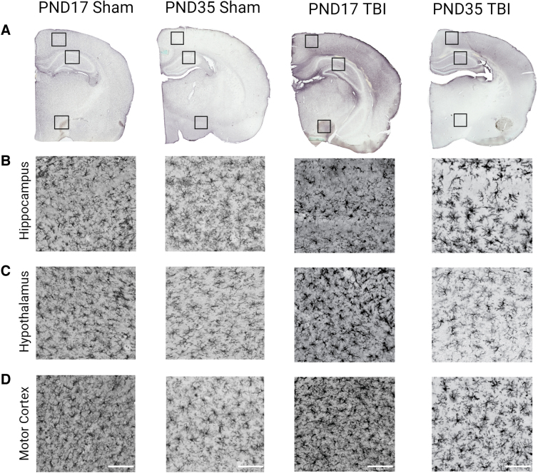
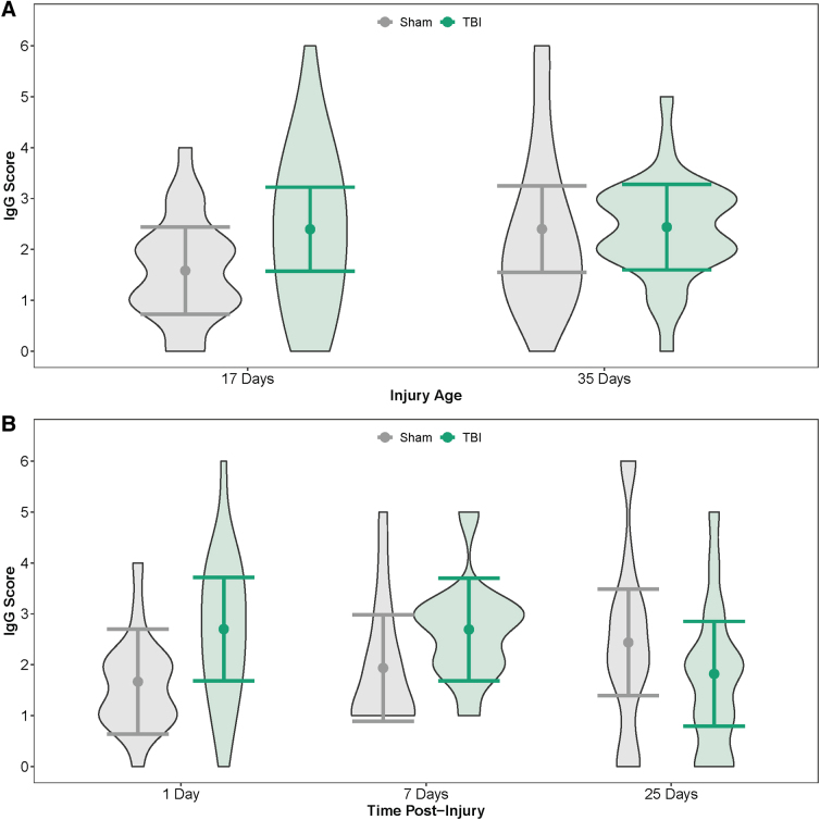
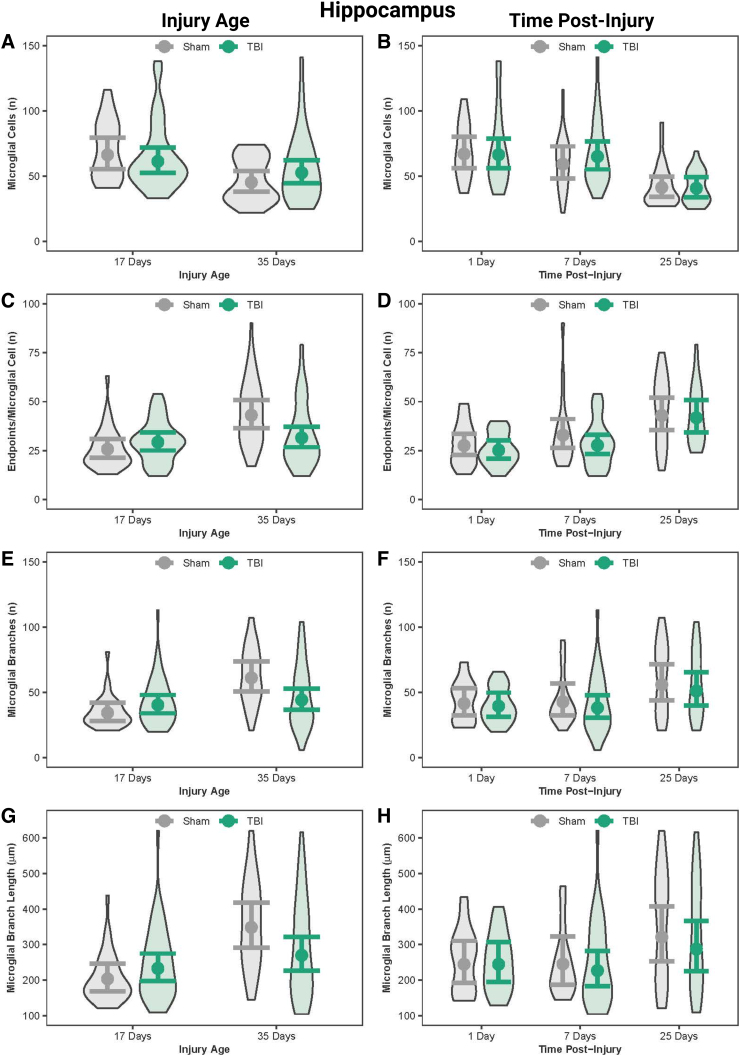
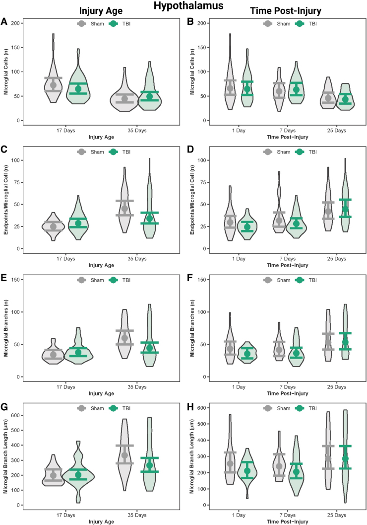
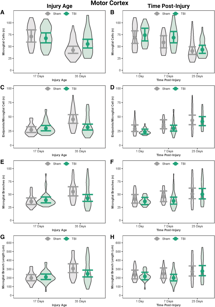
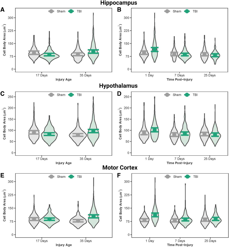
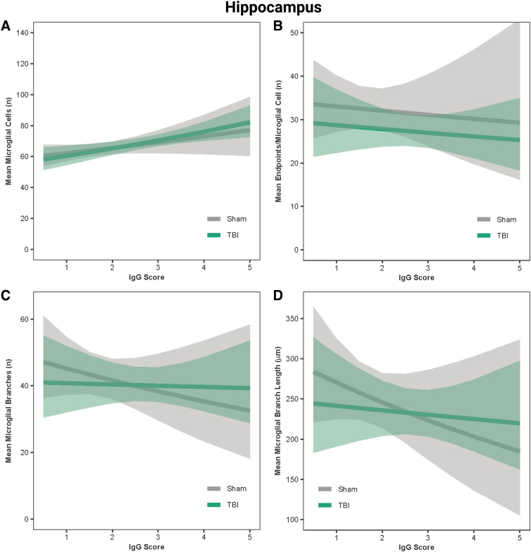
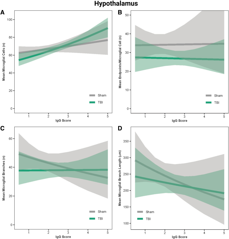
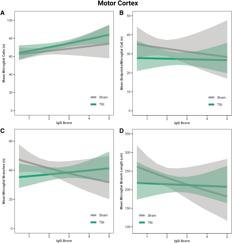
Traumatic brain injury (TBI) disrupts the blood-brain barrier (BBB), which may exacerbate neuroinflammation post-injury. Few translational studies have examined BBB dysfunction and subsequent neuroinflammation post-TBI in juveniles. We hypothesized that BBB dysfunction positively predicts microglial activation and that vulnerability to BBB dysfunction and associated neuroinflammation are dependent on age at injury. Post-natal day (PND)17 and PND35 rats (n = 56) received midline fluid percussion injury or sham surgery, and immunoglobulin-G (IgG) stain was quantified as a marker of extravasated blood in the brain and BBB dysfunction. We investigated BBB dysfunction and the microglial response in the hippocampus, hypothalamus, and motor cortex relative to age at injury and days post-injury (DPI; 1, 7, and 25). We measured the morphologies of ionized calcium-binding adaptor molecule 1-labeled microglia using cell body area and perimeter, microglial branch number and length, end-points/microglial cell, and number of microglia. Data were analyzed using generalized hierarchical models. In PND17 rats, TBI increased levels of IgG compared to shams. Independent of age at injury, IgG in TBI rats was higher at 1 and 7 DPI, but resolved by 25 DPI. TBI activated microglia (more cells and fewer end-points) in PND35 rats compared to respective shams. Independent of age at injury, TBI induced morphological changes indicative of microglial activation, which resolved by 25 DPI. TBI rats had fewer cells and end-points per cell at 1 and 7 DPI than 25 DPI. Independent of TBI, PND17 rats had larger, more activated microglia than PND35 rats; PND17 TBI rats had larger cell body areas and perimeters than PND35 TBI rats. Importantly, we found support in both ages that IgG quantification predicted microglial activation after TBI. The number of microglia increased with increasing IgG, whereas branch length decreased with increasing IgG, which together indicate microglial activation. Our results suggest that stabilization of the BBB after pediatric TBI may be an important therapeutic strategy to limit neuroinflammation and promote recovery.

Keywords: concussion; neuroinflammation; pediatric; vasculature.


Conflict of interest statement

No competing financial interests exist.

Figures

FIG. 1.
FIG. 1.
IgG and Iba1 staining in PND17 and PND35 rats at 1 DPI. (A) IgG staining in PND17 sham, PND35 sham, PND17 TBI, and PND35 TBI 1 day after mFPI or control sham surgery. Iba1 staining in the (B) hippocampus, (C) hypothalamus, and (D) motor cortex of PND17 sham, PND35 sham, PND17 TBI, and PND35 TBI 1 day after mFPI or control sham surgery. Scale bar = 100 μm. DPI, days post-injury; Iba1, ionized calcium-binding adaptor molecule 1; IgG, immunoglobulin-G; mFPI, midline fluid percussion injury; PND, post-natal day; TBI, traumatic brain injury.
FIG. 2.
FIG. 2.
Amount of IgG in the brain by injury age and time post-injury. (A) Predicted IgG scores of the amount of IgG in the brain at PND17 and PND35. (B) Predicted IgG scores of the amount of IgG in the brain across time post-injury (injury ages combined). Results presented as the estimated conditional effects point estimates (dots) and their corresponding 95% confidence intervals (error bars) from generalized linear mixed-effects models. Distributions of the raw data are represented by the background violins. IgG, immunoglobulin-G; PND, post-natal day; TBI, traumatic brain injury.
FIG. 3.
FIG. 3.
Microglial morphology in the hippocampus by injury age and time post-injury. (A) Predicted number of microglial cells across age at injury and (B) time post-injury. (C) Predicted number of end-points per microglia across age at injury and (D) time post-injury. (E) Predicted number of processes per microglia across age at injury and (F) time post-injury. (G) Predicted branch length per microglia across age at injury and (H) time post-injury. Results presented as the estimated conditional effects point estimates (dots) and their corresponding 95% confidence intervals (error bars) from generalized linear mixed-effects models. Distributions of the raw data are represented by the background violins. TBI, traumatic brain injury.
FIG. 4.
FIG. 4.
Microglial morphology in the hypothalamus by injury age and time post-injury. (A) Predicted number of microglial cells across age at injury and (B) time post-injury. (C) Predicted number of end-points per microglia across age at injury and (D) time post-injury. (E) Predicted number of processes per microglia across age at injury and (F) time post-injury. (G) Predicted branch length per microglia across age at injury and (H) time post-injury. Results presented as the estimated conditional effects point estimates (dots) and their corresponding 95% confidence intervals (error bars) from generalized linear mixed-effects models. Distributions of the raw data are represented by the background violins. TBI, traumatic brain injury.
FIG. 5.
FIG. 5.
Microglial morphology in the motor cortex by injury age and time post-injury. (A) Predicted number of microglial cells across age at injury and (B) time post-injury. (C) Predicted number of end-points per microglia across age at injury and (D) time post-injury. (E) Predicted number of processes per microglia across age at injury and (F) time post-injury. (G) Predicted branch length per microglia across age at injury and (H) time post-injury. Results presented as the estimated conditional effects point estimates (dots) and their corresponding 95% confidence intervals (error bars) from generalized linear mixed-effects models. Distributions of the raw data are represented by the background violins. TBI, traumatic brain injury.
FIG. 6.
FIG. 6.
Microglial cell body area in the hippocampus, hypothalamus, and motor cortex. (A) Predicted microglial cell body area across age at injury and (B) time post-injury in the hippocampus. (C) Predicted microglial cell body area across age at injury and (D) time post-injury in the hypothalamus. (E) Predicted microglial cell body area across age at injury in the motor cortex and (F) across time post-injury in the motor cortex. Results presented as the estimated conditional effects point estimates (dots) and their corresponding 95% confidence intervals (error bars) from generalized linear mixed-effects models. Distributions of the raw data are represented by the background violins. TBI, traumatic brain injury.
FIG. 7.
FIG. 7.
IgG score predicted microglial activation in the hippocampus. (A) Predicted relationship between IgG score and the mean number of microglial cells per image. (B) Predicted relationship between IgG score and the mean number of end-points. (C) Predicted relationship between IgG score and the mean number of microglial branches. (D) Predicted relationship between IgG score and the mean microglial branch length per microglial cell. Results presented as the estimated conditional effects point estimates (solid lines) and their corresponding 95% confidence intervals (shaded regions) from generalized linear mixed-effects models. IgG, immunoglobulin-G; TBI, traumatic brain injury.
FIG. 8.
FIG. 8.
IgG score predicted microglial activation in the hypothalamus. (A) Predicted relationship between IgG score and the mean number of microglial cells per image. (B) Predicted relationship between IgG score and the mean number of end-points. (C) Predicted relationship between IgG score and the mean number of microglial branches. (D) Predicted relationship between IgG score and the mean microglial branch length per microglial cell. Results presented as the estimated conditional effects point estimates (solid lines) and their corresponding 95% confidence intervals (shaded regions) from generalized linear mixed-effects models. IgG, immunoglobulin-G; TBI, traumatic brain injury.
FIG. 9.
FIG. 9.
IgG score predicted microglial activation in the motor cortex. (A) Predicted relationship between IgG score and the mean number of microglial cells per image. (B) Predicted relationship between IgG score and the mean number of end-points. (C) Predicted relationship between IgG score and the mean number of microglial branches. (D) Predicted relationship between IgG score and the mean microglial branch length per microglial cell. Results presented as the estimated conditional effects point estimates (solid lines) and their corresponding 95% confidence intervals (shaded regions) from generalized linear mixed-effects models. IgG, immunoglobulin-G; TBI, traumatic brain injury.

References

    1. Serpa RO, Ferguson L, Larson C, et al. . Pathophysiology of pediatric traumatic brain injury. Front Neurol 2021;12:696510; doi: 10.3389/fneur.2021.696510 - DOI - PMC - PubMed
    1. Anderson V, Catroppa C, Morse S, et al. . Functional plasticity or vulnerability after early brain injury? Pediatrics 2005;116(6):1374–1382; doi: 10.1542/peds.2004-1728 - DOI - PubMed
    1. Thurgur H, Pinteaux E. Microglia in the neurovascular unit: blood-brain barrier-microglia interactions after central nervous system disorders. Neuroscience 2019;405:55–67; doi: 10.1016/j.neuroscience.2018.06.046 - DOI - PubMed
    1. Monson KL, Converse MI, Manley GT. Cerebral blood vessel damage in traumatic brain injury. Clin Biomech (Bristol, Avon) 2019;64:98–113; doi: 10.1016/j.clinbiomech.2018.02.011 - DOI - PubMed
    1. Golding EM. Sequelae following traumatic brain injury. The cerebrovascular perspective. Brain Res Brain Res Rev 2002;38(3):377–388; doi: 10.1016/s0165-0173(02)00141-8 - DOI - PubMed

LinkOut - more resources