. 2023 Sep-Dec;18(3):174-180.
doi: 10.5005/jp-journals-10080-1596.

Anterior Hemiepiphysiodesis of the Distal Tibia: A Step-by-step Surgical Technique Guide

Affiliations

Anterior Hemiepiphysiodesis of the Distal Tibia: A Step-by-step Surgical Technique Guide

Alan Katz et al. Strategies Trauma Limb Reconstr. 2023 Sep-Dec.

Abstract

Aim: This paper aims to serve as a guide for surgeons to prepare, execute, and perfect anterior hemiepiphysiodesis of the distal tibia (AHDT).

Background: Treatment of persistent or recurrent equinus deformity following multiple conservative and surgical interventions in patients with idiopathic clubfoot or neuromuscular conditions can be challenging, and multiple surgical options are presented in the existing literature. Anterior hemiepiphysiodesis of the distal tibia is an option that seems to be safe and efficient in treating this entity. To the best of our knowledge, there is not yet any detailed description of this surgical technique in the English literature.

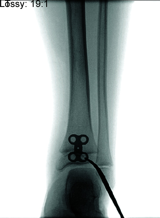
Technique: The AHDT detailed surgical technique includes patient positioning, careful distal anterior tibial approach, placement of guided growth plates, fixation with epiphyseal and metaphyseal screws under fluoroscopic guidance, meticulous closure, and postoperative measures.

Conclusion: This guide can be used pre-operatively to plan the surgery, intra-operatively to aid in smooth and safe step progression, and post-operatively to assist in critical critiquing.

Clinical significance: By understanding the various stages of the surgery as well as the anatomy, pitfalls can be avoided and AHDT can be performed efficiently.

How to cite this article: Katz A, Dumas É, Hamdy R. Anterior Hemiepiphysiodesis of the Distal Tibia: A Step-by-step Surgical Technique Guide. Strategies Trauma Limb Reconstr 2023;18(3):174-180.

Keywords: Ankle; Clubfoot; Equines; Guided growth; Surgical anatomy; Tibia.


Conflict of interest statement

Source of support: Nil Conflict of interest: None

Figures

Fig. 1
Fig. 1
Patient's leg positioning, including leg elevator and tourniquet
Fig. 2
Fig. 2
Sterile draping
Figs 3A1 to D2
Figs 3A1 to D2
(A1 and A2) Marking the joint line. (A1) Clinical photo; (A2) Under fluoroscopy; (B1 and B2) Marking the physis. (B1) Clinical photo; (B2) Under fluoroscopy; (C1 and C2) Marking the medial border of the distal tibia. (C1) Clinical photo; (C2) Under fluoroscopy; (D1 and D2) Marking the lateral border of the distal tibia. (D1) Clinical photo; (D2) Under fluoroscopy
Fig. 4
Fig. 4
Marking the incision
Fig. 5
Fig. 5
Extensor retinaculum incision in line with skin incision, lateral to tibialis anterior tendon. Tibialis anterior tendon is exposed
Fig. 6
Fig. 6
Identification of tibialis anterior in self-retaining retractor and extensor hallucis longus (EHL) in hemostat. (Neurovascular bundle is lateral to EHL)
Figs 7A and B
Figs 7A and B
(A) Incision of the fascia deep to the tibialis anterior tendon; (B) Exposure of the anterior surface of distal tibia
Fig. 8
Fig. 8
Positioning of the first 2-holed plate on the more lateral side of anterior distal tibia under fluoroscopy guidance. Note that the epiphyseal hole is entirely in the epiphysis, neither violating the joint nor the physis
Fig. 9
Fig. 9
Example of the “I” plate (4-holed) that is too large for this patient, with the screw hole overlapping with the physis
Figs 10A and B
Figs 10A and B
(A) Securing plates with central k-wires in the physis; (B) Under fluoroscopic guidance making sure to be centred on the AP
Fig. 11
Fig. 11
Placing k-wires in plate holes guiding for screw placement. Note the plate is distalised to assure distal screw is entirely in the epiphysis, avoiding the physis
Figs 12A and B
Figs 12A and B
Screw placement over the k-wires. (A) Epiphyseal screw placement first, over k-wire; (B) Plate fixation with screws and k-wires out
Fig. 13
Fig. 13
K-wires placement for the second plate, parallel to the screws of the first plate
Fig. 14
Fig. 14
Plates fixated with screws
Figs 15A and B
Figs 15A and B
(A) Fluoroscopy in full plantar flexion; (B) Full dorsiflexion, making sure the plate does not impinge with full ROM
Figs 16A and B
Figs 16A and B
Final fluoroscopy (A) AP; (B) Lateral
Figs 17A to C
Figs 17A to C
(A) Closure of the fascia deep to the tibialis anterior tendon over the plates; (B) The fascia closed; (C) Tendons in normal anatomical position over the deep fascia
Figs 18A and B
Figs 18A and B
(A) Closure of extensor retinaculum over the tendons; (B) Extensor retinaculum closed
Fig. 19
Fig. 19
Closure of the skin

References

    1. Ebert N, Ballhause TM, Babin K, et al. Correction of recurrent equinus deformity in surgically treated clubfeet by anterior distal tibial hemiepiphysiodesis. J Pediatr Orthop. 2020;40(9):520–525. doi: 10.1097/BPO.0000000000001609. - DOI - PubMed
    1. Al-Aubaidi Z, Lundgaard B, Pedersen NW. Anterior distal tibial epiphysiodesis for the treatment of recurrent equinus deformity after surgical treatment of clubfeet. J Pediatr Orthop. 2011;31(6):716–720. doi: 10.1097/BPO.0b013e31822109b6. - DOI - PubMed
    1. Giertych BF, Galli SH, Halanski MA, et al. “Anterior hemi-epiphysiodesis of the distal tibia for residual equinus deformity in children with clubfeet.”. Journal of the Pediatric Orthopaedic Society of North America. 2022;4(1):1–11. doi: 10.55275/JPOSNA-2022-0004. - DOI
    1. Zargarbashi R, Abdi R, Bozorgmanesh M, et al. Anterior distal hemiepiphysiodesis of tibia for treatment of recurrent equinus deformity due to Flat-Top talus in surgically treated clubfoot. J Foot Ankle Surg. 2020;59(2):418–422. doi: 10.1053/j.jfas.2019.08.018. - DOI - PubMed
    1. Mishra AS, Shrestha J, Rajan RA. Anterior distal tibial guided growth for recurrent equinus deformity in idiopathic congenital talipes equinovarus treated with the ponseti method. Foot Ankle Surg. 2023;29(4):355–360. doi: 10.1016/j.fas.2023.03.006. - DOI - PubMed

LinkOut - more resources