Review
. 2023 Sep 30;2023(4):e202328.
doi: 10.21542/gcsp.2023.28.

Myocarditis and COVID-19 related issues

Affiliations
Review

Myocarditis and COVID-19 related issues

Michele Ciabatti et al. Glob Cardiol Sci Pract. .

Abstract

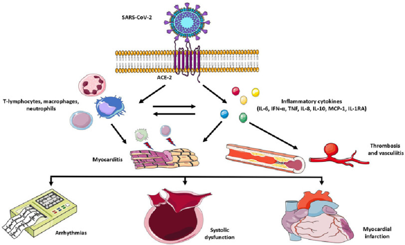
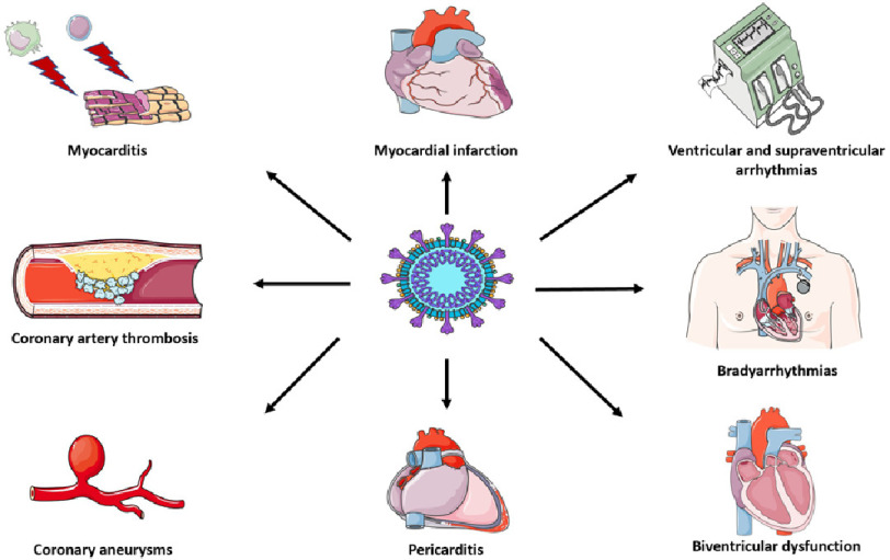
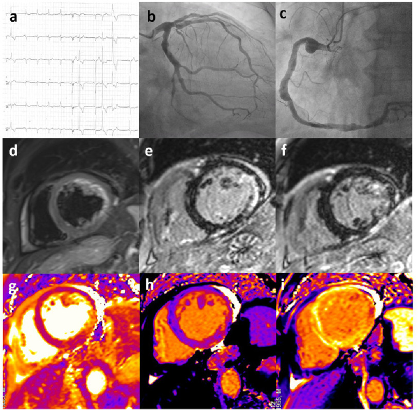
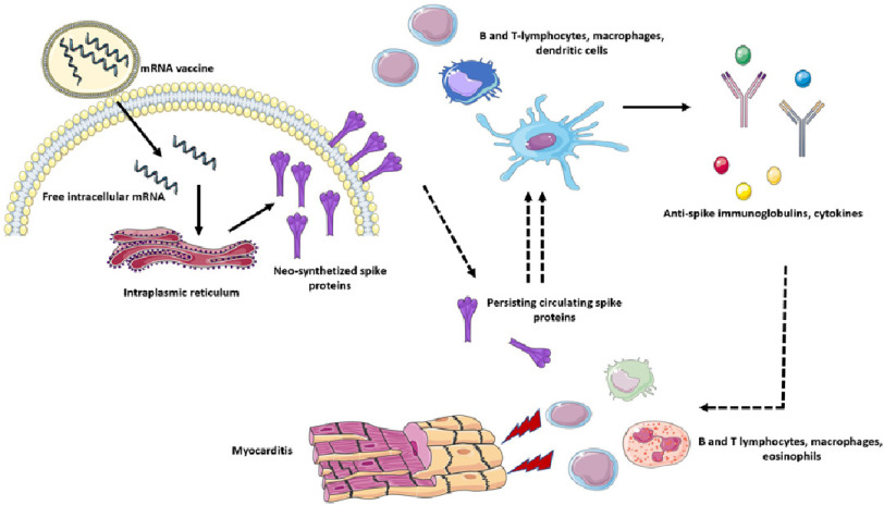
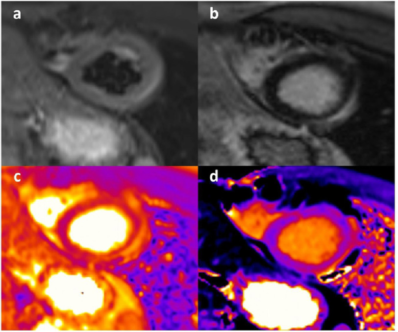
The recent COVID-19 (Coronavirus Disease 2019) pandemic by SARS-CoV2 infection has caused millions of deaths and hospitalizations across the globe. In the early pandemic phases, the infection had been initially considered a primary pulmonary disease. However, increasing evidence has demonstrated a wide range of possible cardiac involvement. Most of systemic and cardiac damage is likely sustained by a complex interplay between inflammatory, immune-related and thrombotic mechanisms. Biventricular failure and myocardial damage with elevation of cardiac biomarkers have been reported in COVID-19 patients, although histological demonstration of acute myocarditis has been rarely documented. Indeed while cardiac magnetic resonance findings include different patterns of myocardial involvement in terms of late gadolinium enhancement, histological data from necropsy and endomyocardial biopsy showed peculiar inflammatory patterns, mostly composed by macrophages. On the other hand COVID-19 vaccines based on mRN technology have been also associated with increased risk of myocarditis. COVID-19 and mRNA vaccine-related myocarditis present different clinical and imaging presentations and recent data suggest the presence of distinctive immunological mechanisms involved.


Figures

Figure 1.
Figure 1.. Mechanisms of cardiovascular complications of SARS-CoV2 infection.
SARS-CoV-2 enters the cellular membrane due to its affinity to ACE-2 as a receptor. The viral infection determines a marked dysregulated innate immune response with important cellular (T-lymphocytes, macrophages, neutrophils) and cytokine (IL-6, IFN-α, TNF, IL-8, IL-10, MCP-1, IL-1RA) activation. These immune and inflammatory processes can provoke myocarditis, thrombosis, and vasculitis in the myocardium. Arrhythmias, biventricular dysfunction and acute coronary syndromes can occur in these patients. Key: SARS-CoV-2, severe acute respiratory syndrome coronavirus 2; IL-6, interleukin 6; IFN-α, interferon alpha; TNF, tumor necrosis factor; IL-8, interleukin 8; IL-10 interleukin 10; MCP-1, Monocyte chemoattractant protein-1; IL-1RA, interleukin 1 receptor antagonist.
Figure 2.
Figure 2.. Cardiovascular manifestations of SARS-CoV2 infection.
SARS-CoV-2 mainly causes macrophage and lymphocytic myocarditis, while its direct pathogenetic role is still debated. Due to its peculiar vascular tropism, many cases of arterial and venous thrombosis and coronary vasculitis have been described in literature. These pathogenetic mechanisms can lead to biventricular dysfunction, brady and tachyarrhythmias and myocardial infarction (even with non-obstructive coronary arteries). Pericarditis is another possible complication of COVID-19 infection. Key: COVID-19, coronavirus disease 2019; SARS-CoV-2, severe acute respiratory syndrome coronavirus 2.
Figure 3.
Figure 3.. A case of COVID-19-related cardiovascular damage.
A 65-year-old male presenting with severe left ventricular dysfunction and non-sustained ventricular tachycardia shortly after hospitalization due to COVID-19 pneumonia. (a) ECG at presentation showed the presence of sinus rhythm with normal atrioventricular conduction, left anterior hemiblock, QRS fragmentation in V1–V2 leads and negative lateral T waves; polymorphic ventricular beats were present. (b–c) Coronary angiography showed diffuse aneurysmatic lesions in the absence of critical stenosis in the circumflex (b) and right (c) coronary artery. (d) CMR demonstrated the presence of myocardial oedema in the LV inferolateral walls on T2-weighted images (short-tau inversion recovery, STIR) with mild pericardial effusion. (e–f) Late sequences after contrast administration showed the presence of extensive ischaemic LGE in the mid LV lateral wall and midwall LGE in the apical anterolateral wall. LGE was detected also in the anterior RV wall. (g) T2 mapping confirmed the presence of inflammation in the lateral LV wall. h and i: Image analysis of Native and post-contrast T1 mapping (ShMOLLI at 1.5 T) showed increased T1 and ECV values matching LGE sequences in the inferior and lateral wall. Key: COVID-19, coronavirus disease 2019; CMR, cardiac magnetic resonance; LV, left ventricle; STIR, short-tau inversion recovery; LGE, late gadolinium enhancement; RV, right ventricle; ECV, extracellular volume.
Figure 4.
Figure 4.. Proposed mechanisms of COVID-19 mRNA vaccine-related myocarditis.
COVID-19 vaccines based on mRNA technology enter cells by endocytosis and release their mRNA (coding for COVID-19 spike protein) content in the cytoplasm. The mRNA is therefore transcripted in the endoplasmic reticulum into numerous spike proteins. These molecules are then exposed in the outer cellular membrane and presented to B and T-lymphocytes, dendritic cells, dedicated macrophages and other antigen presenting cells. The antigen exposure and recognition lead to marked immune cells activation and expansion with consequent immunoglobulin and cytokine production. It has been proposed that persistent free circulating spike proteins could determine an excessive and dysregulated adaptive immune response. These immune pathways could therefore play a role in determining myocarditis with extensive lymphocytes, macrophages and eosinophilic infiltrates. Key: COVID-19, coronavirus disease 2019; mRNA, messenger ribonucleic acid.
Figure 5.
Figure 5.. A case of CIVID-19 mRNA vaccine-related acute myocarditis.
A 23-year-old male presenting with chest pain and elevation of markers of cardiac damage 3 days after the administration of a second dose of mRNA vaccine. (a) STIR sequences showed presence of subepicardial oedema in the mid-apical wall. (b) late sequences after contrast demonstrated corresponding LGE in the same segments. (c) T2 mapping sequences confirmed the presence of inflammation in the lateral wall. (d) presence of increased T1 mapping values in the corresponding segments on native T1 mapping sequences. Key: mRNA, messenger ribonucleic acid; STIR, short-tau inversion recovery; LGE, late gadolinium enhancement.

Similar articles

Cited by

References

    1. Shi S, Qin M, Shen B, Cai Y, Liu T, Yang F, Gong W, Liu X, Liang J, Zhao Q, Huang H, Yang B, Huang C. Association of cardiac injury with mortality in hospitalized patients with COVID-19 in Wuhan, China. JAMA Cardiol. 2020;5(7):802–810. - PMC - PubMed
    1. Zhou F, Yu T, Du R, Fan G, Liu Y, Liu Z, Xiang J, Wang Y, Song B, Gu X, Guan L, Wei Y, Li H, Wu X, Xu J, Tu S, Zhang Y, Chen H, Cao B. Clinical course and risk factors for mortality of adult inpatients with COVID-19 in Wuhan, China: a retrospective cohort study. Lancet. 2020;395(10229):1054–1062. - PMC - PubMed
    1. Zhang L, Yan X, Fan Q, Liu H, Liu X, Liu Z, Zhang Z. D-dimer levels on admission to predict in-hospital mortality in patients with Covid-19. J Thromb Haemost. 2020;18(6):1324–1329. - PMC - PubMed
    1. Kotecha T, Knight DS, Razvi Y, Kumar K, Vimalesvaran K, Thornton G, Patel R, Chacko L, Brown JT, Coyle C, Leith D, Shetye A, Ariff B, Bell R, Captur G, Coleman M, Goldring J, Gopalan D, Heightman M, Hillman T, Howard L, Jacobs M, Jeetley PS, Kanagaratnam P, Kon OM, Lamb LE, Manisty CH, Mathurdas P, Mayet J, Negus R, Patel N, Pierce I, Russell G, Wolff A, Xue H, Kellman P, Moon JC, Treibel TA, Cole GD, Fontana M. Patterns of myocardial injury in recovered troponin-positive COVID-19 patients assessed by cardiovascular magnetic resonance. Eur Heart J. 2021;42(19):1866–1878. - PMC - PubMed
    1. Tanacli R, Doeblin P, Götze C, Zieschang V, Faragli A, Stehning C, Korosoglou G, Erley J, Weiss J, Berger A, Pröpper F, Steinbeis F, Kühne T, Seidel F, Geisel D, Cannon Walter-Rittel T, Stawowy P, Witzenrath M, Klingel K, Van Linthout S, Pieske B, Tschöpe C, Kelle S. COVID-19 vs. classical myocarditis associated myocardial injury evaluated by cardiac magnetic resonance and endomyocardial biopsy. Front Cardiovasc Med. 2021;8:737257. - PMC - PubMed

LinkOut - more resources