. 2024 Feb 19:35:477-494.
doi: 10.1016/j.bioactmat.2024.02.004. eCollection 2024 May.

Urchin-like multiscale structured fluorinated hydroxyapatite as versatile filler for caries restoration dental resin composites

Affiliations

Urchin-like multiscale structured fluorinated hydroxyapatite as versatile filler for caries restoration dental resin composites

Shuning Zhang et al. Bioact Mater. .

Abstract

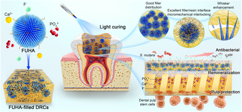
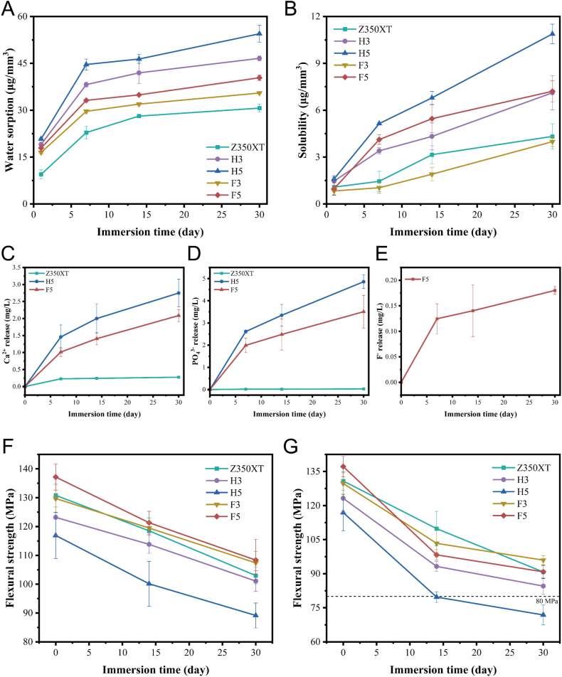
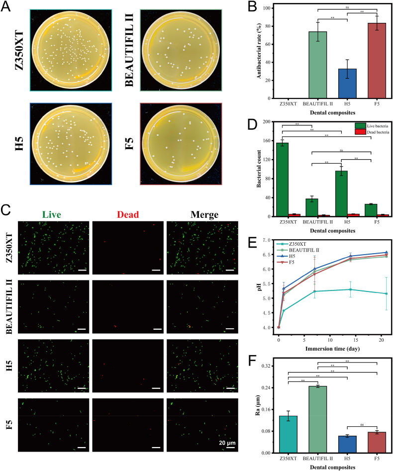
Caries is one of the most prevalent human diseases, resulting from demineralization of tooth hard tissue caused by acids produced from bacteria, and can progress to pulpal inflammation. Filling restoration with dental resin composites (DRCs) is currently the most common treatment for caries. However, existing DRCs suffer from low fracture strength and lack comprehensive anti-caries bioactivity including remineralization, pulp protection, and anti-cariogenic bacteria effects. In this study, inspired by plant roots' ability to stabilize and improve soil, fluorinated urchin-like hydroxyapatite (FUHA) with a three-dimensional whisker structure and bioactive components of calcium, phosphorus, and fluorine was designed and synthesized by a dynamic self-assembly method. Furthermore, versatile FUHA particles with different loading fractions were used as functional fillers to fabricate methacrylate-based DRCs, where the urchin-like hydroxyapatite (UHA) filled DRCs and commercial DRCs (Z350XT and BEAUTIFIL II) served as the control groups. The results demonstrated that FUHA with 50 wt% loading in resin matrix endowed DRC (F5) with excellent physicochemical properties, dentin remineralization property, cell viability, promotion of dental pulp stem cells mineralization, and antibacterial properties. Meanwhile, F5 also presented good clinical handling and aesthetic characteristics. Therefore, structure/functional-integrated FUHA filled DRCs have potential as a promising strategy for tooth restoration and anti-caries bioactivity.

Keywords: Anti-caries bioactivity; Dental resin composites; Micromechanical interlocking; Tooth remineralization; Versatile fluorinated hydroxyapatite.


Conflict of interest statement

The authors declare no competing financial interests or personal relationships that could have appeared to influence the work reported in this paper.

Figures

Image 1
Graphical abstract
Scheme 1
Scheme 1
Scheme of FUHA-filled light-curing DRCs for dentin caries restorations.
Fig. 1
Fig. 1
Characterization of UHA, FUHA and their silanization. (A) Flowchart for the synthesis of root-inspired FUHA. (B) FE-SEM images of UHA and FUHA. (C) TEM images and elemental mapping of UHA and FUHA. (D) XPS spectra and contents of F, Ca. P, O in UHA and FUHA. (E) XRD patterns of UHA and FUHA, HA standard card JCPDS: 09–0432 (gray line). (F) FT-IR spectra and (G) TGA curves of UHA, FUHA, silanized UHA, and silanized FUHA.
Fig. 2
Fig. 2
Physicochemical properties of DRCs. Viscosity (A), curing depth (B), DC (C), FS (D), FM (E), WOF (F), KIC (G), CS (H), FE-SEM images of the fracture morphologies (excluding Z350XT), (I) spectral transmittance curves (J) and digital photos of the 1 mm thick disks (K) of Z350XT, H3, H5, F3 and F5. *p < 0.05, **p < 0.001.
Fig. 3
Fig. 3
Performance of DRCs in body-temperature and moist environments. (A) Water sorption and (B) solubility of Z350XT, H3, H5, F3 and F5 for 1, 7, 14, and 30 days. (C) Release of calcium ions and (D) phosphate from Z350XT, H5 and F5 for 7, 14, and 30 days. (E) Release of fluoride ions from F5 for 7, 14, and 30 days. (F) FS of Z350XT, H3, H5, F3 and F5 after soaking in SBF of pH = 7.4 for 0, 14, and 30 days. (G) FS of Z350XT, H3, H5, F3 and F5 after soaking in SBF of pH = 4 for 0, 14, and 30 days.
Fig. 4
Fig. 4
Remineralization of DRCs. (A) FE-SEM images of surface morphologies of Z350XT, H5 and F5 after soaking in SBF for 1, 14, and 30 days. (B) FE-SEM images of dentin slices, demineralized dentin slices, and demineralized dentin slices being treated with Z350XT, H5, and F5 and soaked in SBF for 14 days. (C) EDS spectra of dentin tubules, demineralized dentin tubules, and demineralized dentin tubules treated with Z350XT, H5 and F5 and soaked in SBF for 14 days. (D) Vickers microhardness of dentin slices, demineralized dentin slices, and demineralized dentin slices being treated with Z350XT, H5, and F5 and soaked in SBF for 14 days. (E) Micro-CT images of artificial cavities and artificial cavities filled with Z350XT, H5 and F5 and soaked in SBF for 30 days. (F) Mineral densities in the original demineralized areas of the artificial cavities after being filled with Z350XT, H5, and F5 (boxed in Fig. 5E). *p < 0.05, **p < 0.001.
Fig. 5
Fig. 5
Cell activity of DRCs. (A) Fluorescence microscopy images of live/dead staining of hDPSCs cultured in blank medium and Z350XT, H3, H5, F3, and F5 extracts for 1, 4, and 7 days: green (live) and red (dead). (B) Surface coverage of live and dead hDPSCs from evaluating fluorescent images with ImageJ software. (C) CCK-8 assay of hDPSCs cultured in blank medium and Z350XT, H3, H5, F3, and F5 extracts for 0, 1, 3, 5, and 7 days. (D) Migration assay of hDPSCs cultured in blank medium and Z350XT, H3, H5, F3, and F5 extracts for 24 h *p < 0.05.
Fig. 6
Fig. 6
Mineralized differentiation of hDPSCs in vitro. (A) ALP staining of hDPSCs and (B) semiquantitative analysis of ALP activity in hDPSCs cultured in osteogenic induction medium and Z350XT, H3, H5, F3, and F5 extracts for 14 days. (C) ARS staining of hDPSCs and (D) quantitative analysis of ARS staining of hDPSCs cultured in osteogenic induction medium and Z350XT, H3, H5, F3, and F5 extracts for 21 days. (E–I) The mRNA expression of ALP, OPN and OCN in hDPSCs cultured in osteogenic induction medium and Z350XT, H3, H5, F3, and F5 extracts for 14 days, while the mRNA expression of DMP-1 and DSPP in hDPSCs cultured for 21 days. (J, K) The immunofluorescence staining images of OPN and OCN in hDPSCs cultured in osteogenic induction medium and Z350XT, H5, and F5 extracts for 14 days: blue (DAPI), red (F-actin), green (OPN or OCN). *p < 0.05, **p < 0.001.
Fig. 7
Fig. 7
Antibacterial effects and environment of DRCs. (A) Colonies of S. mutans eluted from the surfaces of Z350XT, BEAUTIFIL Ⅱ, H5, and F5 after incubation. (B) Inhibition of S. mutans adhesion by BEAUTIFIL Ⅱ, H5 and F5 with zero antibacterial rate of Z350XT. (C) Fluorescence microscopy images of live/dead staining of S. mutans cultured in Z350XT, BEAUTIFIL Ⅱ, H5, and F5's cultures for 3 days. (D) Counts of live and dead bacteria using ImageJ software. (E) The solution pH of Z350XT, BEAUTIFIL II, H5, and F5 soaked in SBF at pH = 4 for 1, 7, 14, and 21 days. (F) Roughness of the surface of Z350XT, BEAUTIFIL Ⅱ, H5, and F5 after 20 s of polishing. **p < 0.001.
Fig. 8
Fig. 8
In vivo effects of Z350XT and F5. (A) Demineralized dentin slices were fixed in the rat oral cavity. (B) FE-SEM image of S. mutans on the surface of Z350XT and F5. (C) FE-SEM image of the dentin slices and their longitudinal sections after Z350XT and F5 treatment.

References

    1. Pitts N.B., Zero D.T., Marsh P.D., Ekstrand K., Weintraub J.A., Ramos-Gomez F., Tagami J., Twetman S., Tsakos G., Ismail A. Dental caries. Nat. Rev. Dis. Prim. 2017;3 doi: 10.1038/nrdp.2017.30. - DOI - PubMed
    1. Selwitz R.H., Ismail A.I., Pitts N.B. Dental caries. Lancet. 2007;369:51–59. doi: 10.1016/S0140-6736(07)60031-2. - DOI - PubMed
    1. Xu Y., You Y., Yi L., Wu X., Zhao Y., Yu J., Liu H., Shen Y., Guo J., Huang C. Dental plaque-inspired versatile nanosystem for caries prevention and tooth restoration. Bioact. Mater. 2023;20:418–433. doi: 10.1016/j.bioactmat.2022.06.010. - DOI - PMC - PubMed
    1. Love R.M., Tanner A.C.R. In: Microbiology of Dental Caries and Dentinal Tubule Infection. first ed. Fouad A.F., editor. John Wiley & Sons, Inc.; Hoboken: 2017. pp. 25–49. (Endod. Microbiol.). - DOI
    1. Cheng L., Zhang L., Yue L., Ling J., Fan M., Yang D., Huang Z., Niu Y., Liu J., Zhao J., Li Y., Guo B., Chen Z., Zhou X. Expert consensus on dental caries management. Int. J. Oral Sci. 2022;14:17. doi: 10.1038/s41368-022-00167-3. - DOI - PMC - PubMed

LinkOut - more resources