Case Reports
. 2024 Jan 3;2024(1):e202406.
doi: 10.21542/gcsp.2024.6.

Use of veno-venous extracorporeal membrane oxygenation for stabilization prior to redo sternotomy for aortic pseudoaneurysm repair

Affiliations
Case Reports

Use of veno-venous extracorporeal membrane oxygenation for stabilization prior to redo sternotomy for aortic pseudoaneurysm repair

Anson Y Lee et al. Glob Cardiol Sci Pract. .

Abstract

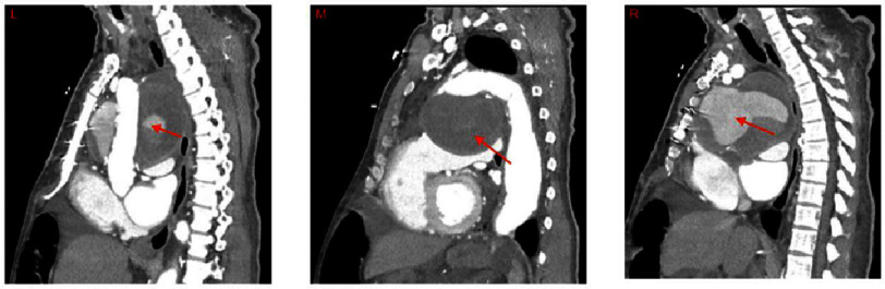
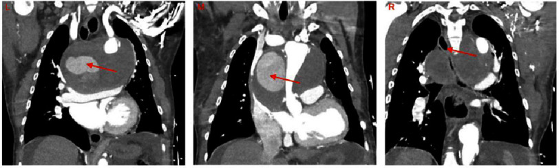
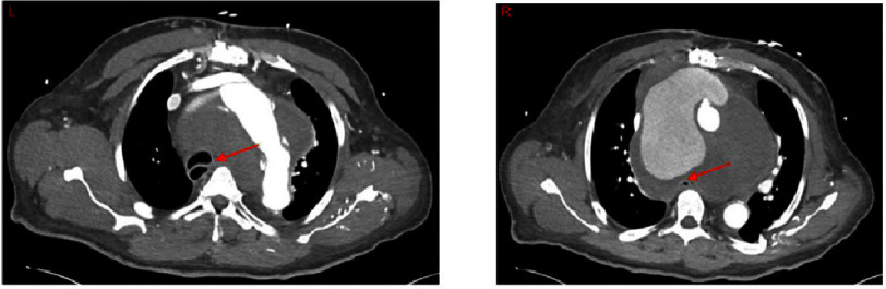
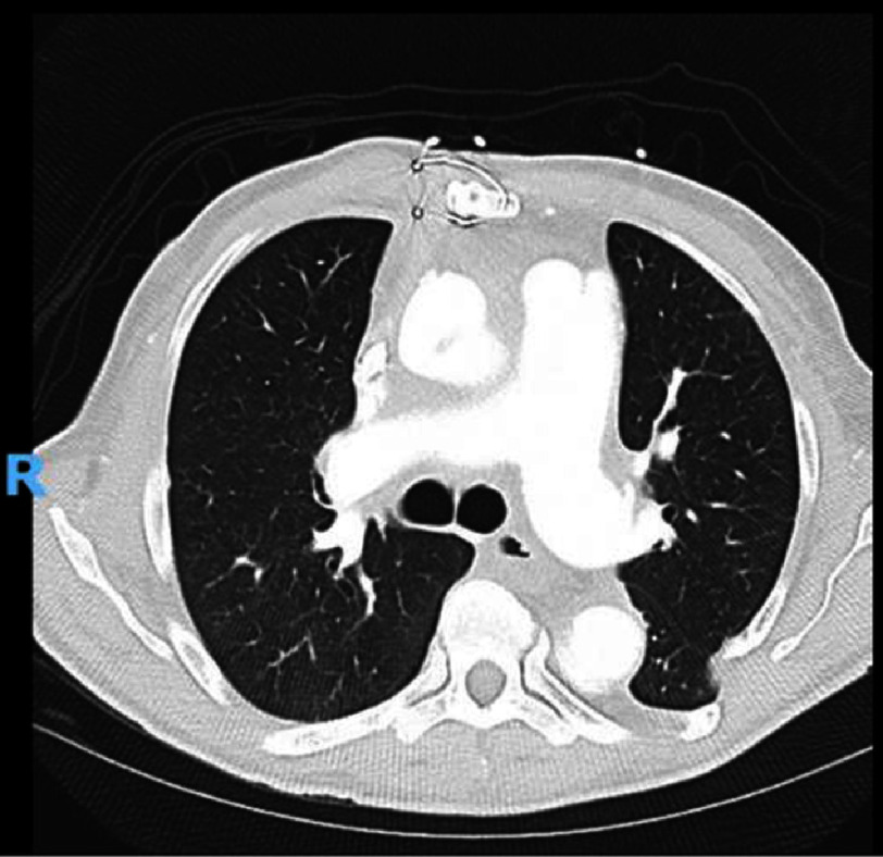
Background: Aortic pseudoaneurysms are particularly dangerous because of the risk of rupture and compression of mediastinal structures, including the trachea, and resultant respiratory distress. If respiratory distress progresses to respiratory failure, extracorporeal membrane oxygenation may be used to provide oxygenation prior to or during pseudoaneurysm repair. Case presentation: A 62-year-old male with a history of emergent aortic ascending and arch replacement for Stanford Type A dissection 10 months prior presented to his primary care physician with dyspnea. Chest radiography revealed a widened mediastinum, and subsequent computed tomography angiogram revealed a pseudoaneurysm at the distal suture line of the aortic arch replacement. Due to the location of the pseudoaneurysm, the patient's trachea was compressed, and he was emergently placed on veno-venous (VV) extracorporeal membrane oxygenation (ECMO) following unsuccessful intubation for respiratory distress. Two days later, the patient underwent a redo sternotomy and repair of a 2-3 mm defect in the anterior aspect of the distal suture line of the prior aortic arch replacement. The patient progressed well and was discharged on postoperative day 13. What we learned: Using a combination of peripheral bypass, hypothermic circulatory arrest, delayed closure, and respiratory support, this case demonstrates how even complex patients can be successfully treated with multiple strategies.


Figures

Figure 1.
Figure 1.. Sagittal Images of CT scan of a patient on VV ECMO with an aortic pseudoaneurysm at the distal suture line of a prior aortic arch replacement.
Left: Large pseudoaneurysm (arrow) behind the aortic graft, compressing the trachea and esophagus posteriorly. Middle: Large pseudoaneurysm below arch (arrow), compressing heart inferiorly. Right: Large pseudoaneurysm (arrow) just posterior to the sternum, compressing the heart inferiorly while compressing the esophagus and trachea posteriorly. Abbreviations: CT - computed tomography, CTA - computed tomography angiogram, VV ECMO - venovenous extracorporeal membrane oxygenation.
Figure 2.
Figure 2.. Coronal Images of CT scan of a patient on VV ECMO with an aortic pseudoaneurysm at the distal suture line of a prior aortic arch replacement.
Left: Large pseudoaneurysm (arrow) with graft anastomosis visible just distal to the left subclavian, left atrium and heart compressed inferiorly. Middle: Large pseudoaneurysm (arrow) leading to compression of the aortic graft. Right: Large pseudoaneurysm compressing the trachea (arrow) with rightward displacement. Abbreviations: CT - computed tomography, CTA - computed tomography angiogram, VV ECMO - veno-venous extracorporeal membrane oxygenation.
Figure 3.
Figure 3.. Axial Images of CT scan of a patient on VV ECMO with an aortic pseudoaneurysm at the distal suture line of a prior aortic arch replacement.
Left: Aortic arch with large pseudoaneurysm and compression and distortion of the trachea (arrow) to the right side. Right: Aortic extravasation of contrast into pseudoaneurysm which can be seen compressing the esophagus (arrow). Abbreviations: CT - computed tomography, CTA - computed tomography angiogram, VV ECMO - venovenous extracorporeal membrane oxygenation.
Figure 4.
Figure 4.. Axial Image of a postoperative CT scan of a patient following the repair of an aortic pseudoaneurysm at the distal suture line of a prior aortic arch replacement on VV ECMO.
Axial image of the chest at the level of T7 in a patient following pseudoaneurysm repair 10 months after ascending and aortic arch replacement for acute type A aortic dissection. Mediastinal anatomy and trachea are consistent with resolution of the large pseudoaneurysm. Abbreviations: CT is computed tomography, VV ECMO is venovenous extracorporeal membrane oxygenation.

Similar articles

References

    1. Razzouk A, Gundry S, Wang N, Heyner R, Sciolaro C, Van Arsdell G, Bansal R, Vyhmeister E, Bailey L. Pseudoaneurysms of the aorta after cardiac surgery or chest trauma. Am Surg. 1993;59(12):818–823. - PubMed
    1. Kresowik TF, Khoury MD, Miller BV, Winniford MD, Shamma AR, Sharp WJ, Blecha MB, Corson JD. A prospective study of the incidence and natural history of femoral vascular complications after percutaneous transluminal coronary angioplasty. J Vasc Surg. 1991;13(2):328–335. - PubMed
    1. Bossone E, Ranieri B, Romano L, Russo V, Barbuto L, Cocchia R, Pezzullo F, Amato C, Vriz O, Di Tommaso L, Iannelli G, Czerny M. Acute aortic syndromes: diagnostic and therapeutic pathways. Heart Fail Clin. 2020;16(3):305–315. doi: 10.1016/j.hfc.2020.03.002. doi: - DOI - PubMed
    1. March KL, Sawada SG, Tarver RD, Kesler KA, Armstrong WF. Current concepts of left ventricular pseudoaneurysm: pathophysiology, therapy, and diagnostic imaging methods. Clin Cardiol. 1989;12(9):531–540. doi: 10.1002/clc.4960120911. doi: - DOI - PubMed
    1. Popovic B, Freysz L, Chometon F, Lemoine J, Elfarra M, Angioi M, Selton-Suty C, de Chillou C, Aliot E. Femoral pseudoaneurysms and current cardiac catheterization: evaluation of risk factors and treatment. Int J Cardiol. 2010;141(1):75–80. doi: 10.1016/j.ijcard.2008.11.111. doi: - DOI - PubMed

Publication types

LinkOut - more resources