

Strontium ranelate retards disc degradation and improves endplate and bone micro-architecture in ovariectomized rats with lumbar fusion induced - Adjacent segment disc degeneration
- PMID: 38404727
- PMCID: PMC10884424
- DOI: 10.1016/j.bonr.2024.101744
Strontium ranelate retards disc degradation and improves endplate and bone micro-architecture in ovariectomized rats with lumbar fusion induced - Adjacent segment disc degeneration
Abstract
Objectives: Adjacent segment disc degeneration (ASDD) is one of the long-term sequelae of spinal fusion, which is more susceptible with osteoporosis. As an anti-osteoporosis drug, strontium ranelate (SR) has been reported to not only regulate bone metabolism but also cartilage matrix formation. However, it is not yet clear whether SR has a reversal or delaying effect on fusion-induced ASDD in a model of osteoporosis.
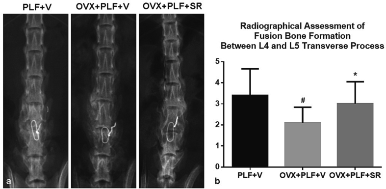
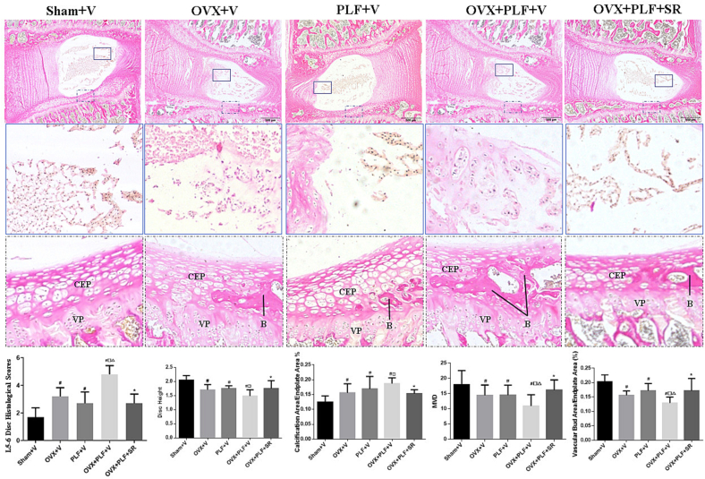
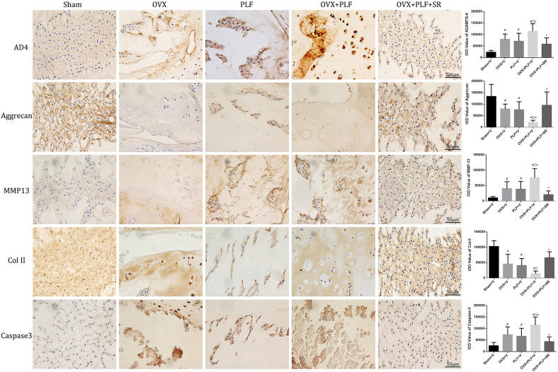
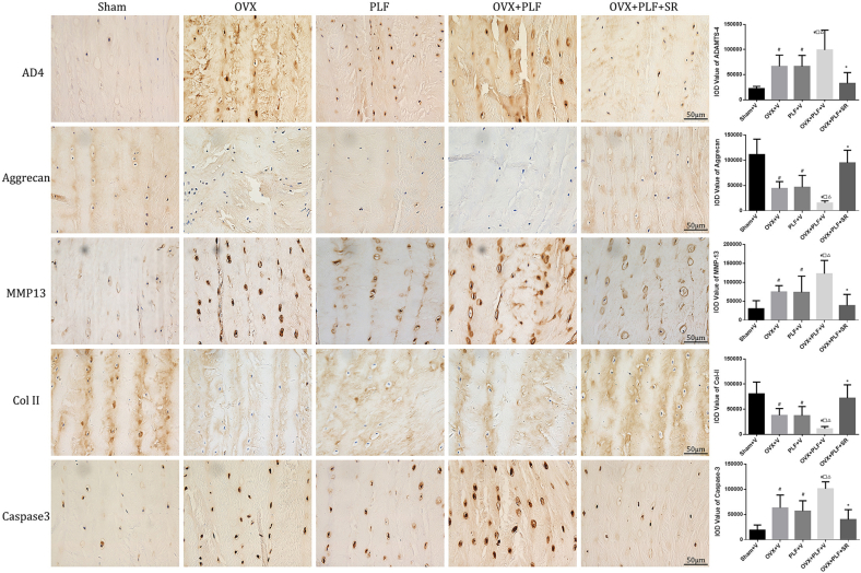
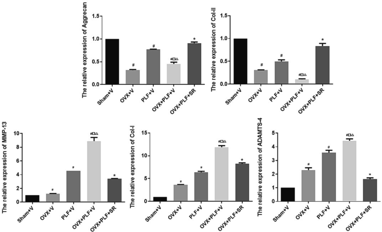
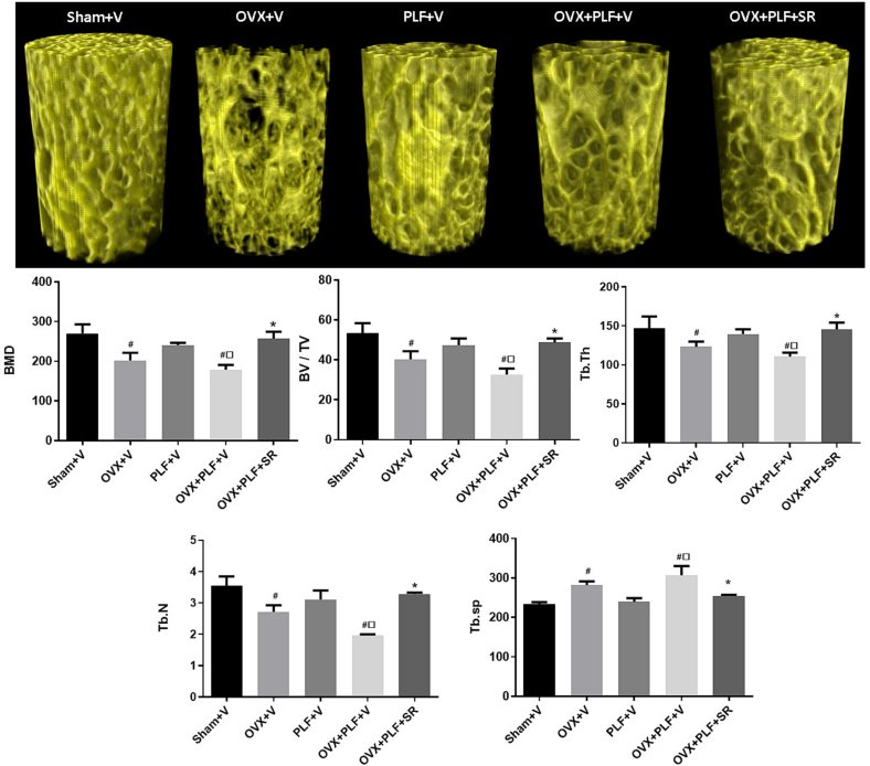
Materials and methods: Fifth three-month-old female Sprague-Dawley rats that underwent L4-L5 posterolateral lumbar fusion (PLF) with spinous-process wire fixation 4 weeks after bilateral ovariectomy (OVX) surgery. Animals were administered vehicle (V) or SR (900 mg/kg/d) orally for 12 weeks post-PLF as follows: Sham+V, OVX + V, PLF + V, OVX + PLF + V, and OVX + PLF + SR. Manual palpation and X-ray were used to evaluate the state of lumbar fusion. Adjacent-segment disc was assessed by histological (VG staining and Scoring), histomorphometry (Disc Height, MVD, Calcification rate and Vascular Bud rate), immunohistochemical (Col-II, Aggrecan, MMP-13, ADAMTS-4 and Caspase-3), and mRNA analysis (Col-I, Col-II, Aggrecan, MMP-13 and ADAMTS-4). Adjacent L6 vertebrae microstructures were evaluated by microcomputed tomography.
Results: Manual palpation and radiographs showed clear evidence of the fused segment's immobility. After 12 weeks of PLF surgery, a fusion-induced ASDD model was established. Low bone mass caused by ovariectomy can significantly exacerbate ASDD progression. SR exerted a protective effect on adjacent segment intervertebral disc with the underlying mechanism possibly being associated with preserving bone mass to prevent spinal instability, maintaining the functional integrity of endplate vascular microstructure, and regulating matrix metabolism in the nucleus pulposus and annulus fibrosus.
Discussion: Anti-osteoporosis medication SR treatments not only maintain bone mass and prevent fractures, but early intervention could also potentially delay degenerative conditions linked to osteoporosis. Taken together, our results suggested that SR might be a promising approach for the intervention of fusion-induced ASDD with osteoporosis.
Keywords: Adjacent segment disc degeneration; Lumbar fusion; Osteoporosis; Ovariectomy; Rat; Strontium Ranelate.
© 2024 The Authors.
Conflict of interest statement
All authors disclosed no relevant relationships.
Figures
Similar articles
-
Alendronate Prevents Intervertebral Disc Degeneration Adjacent to a Lumbar Fusion in Ovariectomized Rats.Spine (Phila Pa 1976). 2015 Oct 15;40(20):E1073-83. doi: 10.1097/BRS.0000000000001092. Spine (Phila Pa 1976). 2015. PMID: 26731708
-
Denosumab alleviates intervertebral disc degeneration adjacent to lumbar fusion by inhibiting endplate osteochondral remodeling and vertebral osteoporosis in ovariectomized rats.Arthritis Res Ther. 2021 May 28;23(1):152. doi: 10.1186/s13075-021-02525-8. Arthritis Res Ther. 2021. PMID: 34049577 Free PMC article.
-
Enhancement of Lumbar Fusion and Alleviation of Adjacent Segment Disc Degeneration by Intermittent PTH(1-34) in Ovariectomized Rats.J Bone Miner Res. 2016 Apr;31(4):828-38. doi: 10.1002/jbmr.2736. Epub 2015 Nov 27. J Bone Miner Res. 2016. PMID: 26542457
-
Protective effect of calcitonin on lumbar fusion-induced adjacent-segment disc degeneration in ovariectomized rat.BMC Musculoskelet Disord. 2015 Nov 9;16:342. doi: 10.1186/s12891-015-0788-7. BMC Musculoskelet Disord. 2015. PMID: 26552386 Free PMC article.
-
Strontium ranelate in osteoporosis.Curr Pharm Des. 2002;8(21):1907-16. doi: 10.2174/1381612023393639. Curr Pharm Des. 2002. PMID: 12171530 Review.
Cited by
-
Co-morbid mechanisms of intervertebral disc degeneration and osteoporosis: biomechanical coupling and molecular pathways synergistically driving degenerative lesions.J Orthop Surg Res. 2025 Jul 14;20(1):652. doi: 10.1186/s13018-025-06075-6. J Orthop Surg Res. 2025. PMID: 40660249 Free PMC article. Review.
References
-
- Abe Y., Takahata M., Ito M., Irie K., Abumi K., Minami A. Enhancement of graft bone healing by intermittent administration of human parathyroid hormone (1-34) in a rat spinal arthrodesis model. Bone. 2007;41-5:775–785. - PubMed
-
- (accessed 4 November /2013).
-
- Benneker L.M., Heini P.F., Alini M., Anderson S.E., Ito K. Young investigator award winner: vertebral endplate marrow contact channel occlusions and intervertebral disc degeneration. Spine (Phila Pa 1976) 2004;2005(30–2):167–173. - PubMed
-
- Bruyere O., Delferriere D., Roux C., Wark J.D., Spector T., Devogelaer J.P., Brixen K., Adami S., Fechtenbaum J., Kolta S., Reginster J.Y. Effects of strontium ranelate on spinal osteoarthritis progression. Ann. Rheum. Dis. 2008;67-3:335–339. - PubMed
LinkOut - more resources
Full Text Sources
Research Materials
